


Recognition of Fuscozetes spitsbergensis (Thor, 1934) n. comb. (Acari, Oribatida, Ceratozetidae), with taxonomic remarks on Fuscozetes
Ermilov, Sergey G.  1
; Makarova, Olga L.
2
; Andrianov, Boris V.
3
and Behan-Pelletier, Valerie M.
4
1
; Makarova, Olga L.
2
; Andrianov, Boris V.
3
and Behan-Pelletier, Valerie M.
4
1✉ University of Tyumen, X-BIO Institute, Tyumen, Russia.
2Severtsov Institute of Ecology and Evolution, Russian Academy of Sciences, Moscow, Russia.
3Vavilov Institute of General Genetics, Russian Academy of Sciences, Moscow, Russia.
4Invertebrate Biodiversity Program, Research Branch, Agriculture and Agri-Food Canada, K.W. Neatby Bldg., Ottawa, Ontario, Canada.
2026 - Volume: 66 Issue: 3 pages: 818-840
https://doi.org/10.24349/812s-xa4jZooBank LSID: D41B17F4-4550-4195-B1B1-3460FB1826D3

Original research
Keywords
Abstract
Introduction
Sig Thor, the Norwegian water mite expert, described Ceratozetes spitsbergensis Thor, 1934 from Large Passage and Magdalene Bay (Magdalenefjorden) on the west coast of Spitsbergen Island, Svalbard, but did not illustrate the species. Subsequently, Behan-Pelletier (1985) redescribed and illustrated this species based on specimens from Svalbard, Brooks Range, Alaska and Pik Aborigen, Magadan Oblast in the Russian Far East. She questioned the placement of this species in Ceratozetes and noted that discovery of juveniles could clarify its relationships. So far, C. spitsbergensis has been recorded from Svalbard (Thor, 1934; Karppinen 1967; Seniczak and Plichta 1978; Lebedeva et al. 2006, 2014; Coulson et al. 2000, 2014, 2015; Ávila‑Jiménez et al. 2019), northern Canada (Behan-Pelletier 1997; Chernov 2004; Behan-Pelletier and Schatz 2010), Mongolia (Bayartogtokh 2010), arctic Russia (Makarova 2002a, 2023; Makarova et al. 2015; Bizin and Makarova 2024), and the Alps in Austrian Tyrol (Fischer et al. 2016). Although the placement of this species was not questioned in any of those publications, a morphological analysis by Behan-Pelletier and Eamer (2009) indicated that C. spitsbergensis was an outgroup to most other Ceratozetes Berlese, 1908 species in any of their equally parsimonious trees, noting that its retention within the genus would need ''further assessment with juvenile and molecular characters''.
Recently, specimens of all ontogenetic instars (adult, larva, proto-, deuto-, tritonymph) of C. spitsbergensis were collected from Taimyr Peninsula, northern Russia. These were morphologically similar to Fuscozetes coulsoni Seniczak and Seniczak, 2020, with a few small differences indicating a possible population level variability as established by Seniczak et al. (2024) for Fuscozetes fuscipes (C.L. Koch, 1844). Our main goal is to combine C. spitsbergensis in the genus Fuscozetes Sellnick, 1928, to redescribe its juvenile instars, and propose F. coulsoni as a junior synonym of F. spitsbergensis n. comb. In addition, we assess the systematic position of F. spitsbergensis, based on mitochondrial COI gene sequences. We provide a revised diagnosis for Fuscozetes, identification keys to the larval and tritonymphal instars in the genus, as well as an update of Fischer et al. (2016) on the distribution and habitats of F. spitsbergensis in the Holarctic region.
Methods
Morphological observation and documentation
For measurement and illustration, specimens were mounted in lactic acid on temporary cavity slides. Body length was measured in lateral view, from the tip of the rostrum to the posterior edge of the gastronotum. Maximum body width refers to the maximum width of the gastronotum in dorsal view. Curved structures, e.g. setae, were measured from the side to avoid foreshortening. All measurements were in micrometers (µm). Formulas for leg setation are given in parentheses according to the sequence trochanter-femur-genu-tibia-tarsus (famulus of tarsus I included). Formulas for leg solenidia are given in square brackets, according to the sequence genu-tibia-tarsus. Paired structures are described in the singular, unless otherwise noted. Drawings were made with a camera lucida using a Leica DM 2500 light microscope. Images were obtained with an AxioCam ICc3 camera using a Carl Zeiss transmission light microscope Axio Lab.A1. For SEM microscopy alcohol-preserved mites were coated with gold and scanned using a TESCAN Mira3 LMU SEM microscope.
Terminology
Morphological terminology used in this paper follows that of Grandjean: see Travé and Vachon (1975) for references, Norton (1977) for leg setal nomenclature, and Norton and Behan-Pelletier (2009) for overview.
Abbreviations
Prodorsum: ro, le, in, bs, ex = rostral, lamellar, interlamellar, bothridial, and exobothridial setae, respectively; ps = prodorsal shield (macrosclerite). Gastronotal region: c1–c3, da, dm, dp, la, lm, lp, h1–h3, p1–p3 = setae; ia, im, ip, ih, ips = cupules; gla = opisthonotal gland opening; oh = humeral organ; δ = ecdysial cleavage line (line of dehiscence); gs = gastronotal shield (macrosclerite), or pygidial sclerite; pls = posterolateral shield (macrosclerite), or opisthonotal gland sclerite; ls = lateral sclerite; vs = ventral sclerite. Gnathosoma: a, m, h = subcapitular setae; or = adoral seta; as = axillary saccule; d, l, cm, acm, ul, su, lt, vt, sup, inf = palp setae; ω = palp solenidion; ep = postpalpal seta; cha, chb = cheliceral setae; Tg = Trägårdh's organ. Epimeral region: 1a, 1b, 1c, 2a, 3a, 3b, 3c, 4a, 4b, 4c = epimeral setae; Cl = Claparède's organ. Anogenital region: g, ag, an, ad = genital, aggenital, anal, and adanal setae, respectively; iad = adanal cupule. Legs: pa = porose area; ω, σ, φ = solenidia; e = famulus; d, l, v, ev, bv, ft, tc, it, p, u, a, s, pv, pl = setae. Instars: LA = larva; PN, DN, TN = proto-, deuto-, and tritonymph, respectively; AD = adult.
Molecular analyses
Nineteen specimens of F. spitsbergensis were analyzed individually. BOLD fragments of the mitochondrial gene COI were obtained according to standard protocols at CCDB (CCDB 2019), using LCO1490/HCO2198 (Folmer et al. 1994) primer pairs. Six new haplotypes were deposited in GenBank. For comparison, we used all full-length BOLD fragments (658 bp in length) of the members of the genus Fuscozetes identified to species level available in GenBank. These sequences belong to Fuscozetes fuscipes (C.L. Koch, 1844), F. pseudosetosus (Shaldybina, 1975), and F. setiger (Trägårdh, 1910). The nucleotide sequence of PCR fragments was determined from the forward and reverse primers on a 3500 Genetic Analyzer using BigDye®Terminator v3.1 Cycle Sequencing Kit reagents (Applied Biosystems, USA) according to the manufacturer's recommendations. Cladistic analyses were performed in MEGA X software (Kumar et al. 2016). Cladograms were constructed using the neighbour-joining method. Substitution model – Maximum Composite Likelihood. Treatment of gaps/missing data – pairwise deletion. Trees are drawn to scale, with branch lengths in units of the number of base substitutions per site. The statistical significance of the resulting clustering of taxa was assessed using bootstrap support. The percentage of replicate trees in which the associated taxa clustered together in the bootstrap test (1000 replicates) is shown next to the branches.
Notes
In addition to the 2025 data (see Material below), a collection of Arctic mites stored in the Synecology Laboratory of the Severtsov Institute of Ecology and Evolution of the Russian Academy of Sciences (Moscow, Russia) was considered in the analysis of the distribution and demography of F. spitsbergensis. Juvenile specimens were associated with adults using criteria outlined by Norton and Ermilov (2014). Specifically, they were found in the same samples and had appropriate size and proportions.
Taxonomy
Family Ceratozetidae
Genus Fuscozetes Sellnick, 1928
Type species Oribata fuscipes C.L. Koch, 1844
Generic diagnosis
Adult — With character states of the Ceratozetidae (Grandjean 1963). Rostrum rounded or undulating, with or without lateral teeth, without medial tooth. Lamellae converging, with long lamellar cusps, with or without lateral and medial teeth. Translamella short, usually wide, sometimes not expressed. Bothridial wall expanded to form well developed ventrolateral and dorsomedial scales, latter extending laterally as free-standing lamina. Genal incision deep, genal tooth well developed. Tutorium narrow, with long cusp, with or without dorsal spines. Pedotectum I large, partially or completely covering acetabulum I. Custodium with short cusp. Discidium lamelliform. Without horizontal folds in integument between and dorsad of acetabula II and III. Notogaster distinctly longer than wide, with comparatively short pteromorphs curved ventrad, without line of desclerotization. Ten to 13 pairs of notogastral setae and four pairs of porose areas present. Six pairs of genital setae. Leg solenidion φ2 arising anterodorsally on tibia I. Femora I and II oval in cross-section, femur II with small to large ventral carina. Solenidion ω2 of tarsus I shorter than ω1 and located posterolateral to ω1.
Juvenile instars — Prodorsal macrosclerite present. Twelve pairs of gastronotal setae in larva, 15 pairs in nymphs. Pair of humeral organs present or absent in larva and nymphs. Gastronotal region with or without many microsclerites in larva; gastronotal region with or without macrosclerite or pygidial sclerite in larva and nymphs. Gastronotal setae borne on macrosclerite without microsclerites at their base; gastronotal setae on unsclerotized integument with or without microsclerites at their base. Humeral sclerite present in nymphs; present or absent in larva. Gastronotal seta c1 inserted on humeral macrosclerite or microsclerite, or on unsclerotized integument, setae c2 and c3 inserted on microsclerites or unsclerotized integument. Leg solenidion ω2 of tarsus I shorter than ω1 and located posterolateral to ω1.
Descriptions
Fuscozetes spitsbergensis (Thor, 1934) n. comb.
(Figures 1–7)
Ceratozetes spitsbergensis in Behan-Pelletier 1985; Behan-Pelletier and Eamer 2009
Ceratozetes cf. spitsbergensis in Bayartogtokh 2010
Ceratozetes sp. near spitsbergensis in Makarova and Behan-Pelletier 2015
Fuscozetes coulsoni Seniczak and Seniczak, 2020 n. syn.
''Sphaerozetes piriformis ? (Nic.) = Oribata piriformis var. setiger Trägårdh, 1910'' in Jørgensen 1934; material studied by M. Hammer [Jørgensen] revised (Makarova and Behan-Pelletier 2015)
? Sphaerozetes piriformis (Nicolet, 1855) in Krivolutsky et al. 1995 (part.)
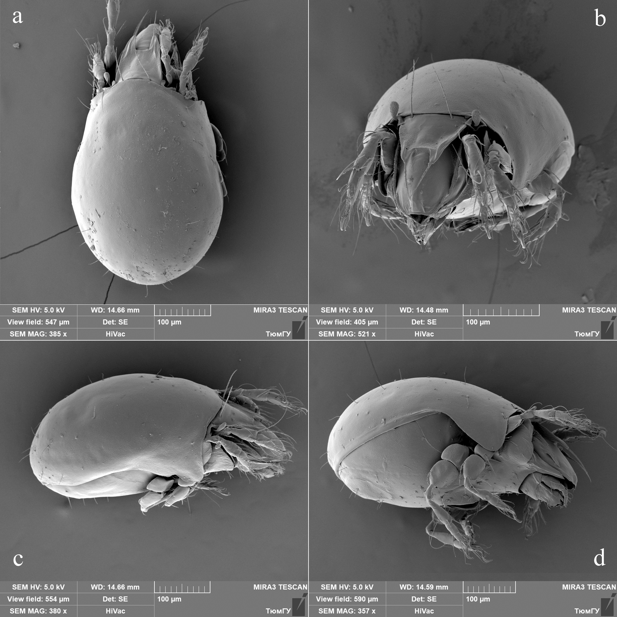
Material — Adults (10 males and 14 females) and juvenile instars (10 larvae, seven protonymphs, eight deutonymphs, and eight tritonymphs): northern Russia, Taimyr, Chelyuskin Peninsula, 77.438724°N, 105.77074°E, 150 m a.s.l., kurums (boulder field, ''stony river''), moss Racomitrium lanuginosum cushion, 9 August 2025 (P. Pavlova). All specimens preserved in a 70% solution of ethanol with a drop of glycerol in the personal collection of the first author.
Additional material: 168 individuals (ind.) of all stages: as above; 19 ind. of all stages: the same district and collector, 77.68079°N, 104.63621°E, 3–5 m a.s.l, Racomitrium lanuginosum cushions, 10 August 2025.
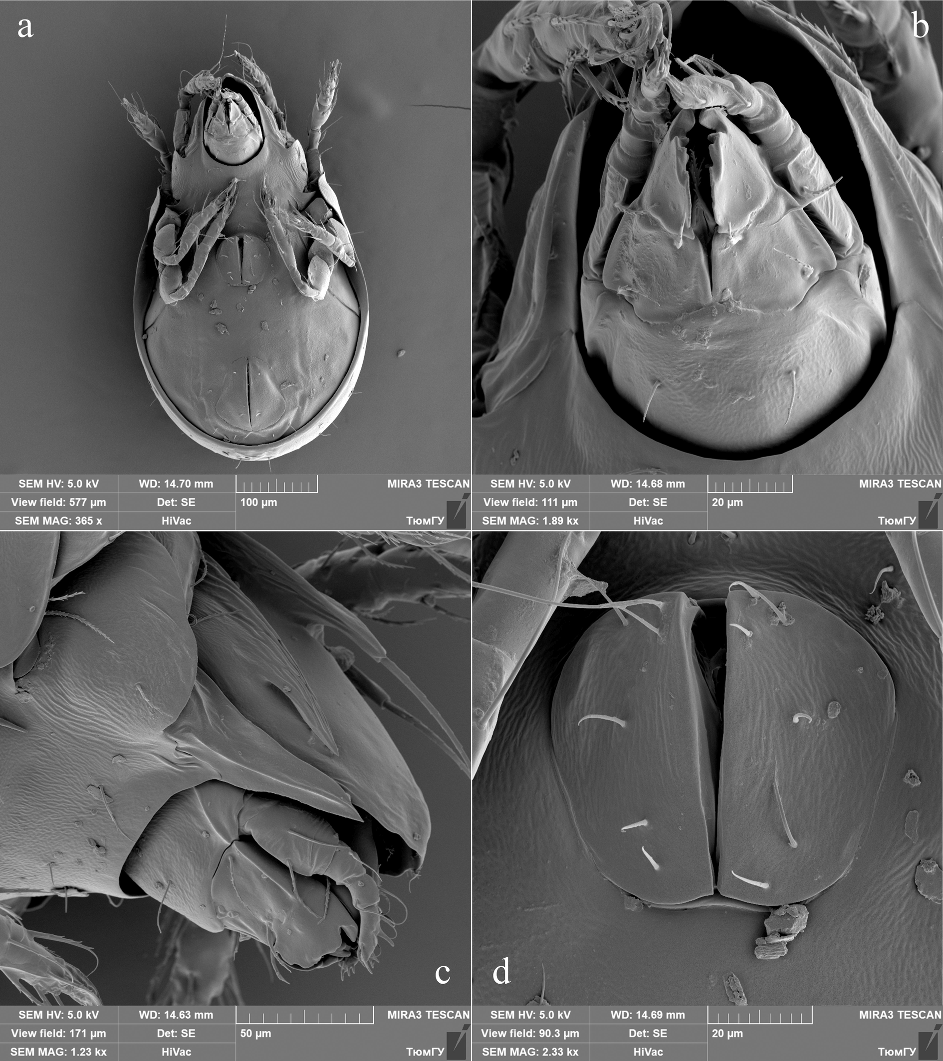
Adult — Adult of this species has been described and figured by Behan-Pelletier (1985), Behan-Pelletier and Eamer (2009), and Seniczak and Seniczak (2020; as F. coulsoni). These latter authors indicated that trochanters III–IV and femur II are flat with a small ventral carina (illustrated in their Fig. 4b, d). The same was illustrated for C. spitsbergensis by Behan-Pelletier (1985, Figures 44–46). However, these carinae are not evident in drawn illustrations of legs of F. coulsoni by Seniczak and Seniczak (2020, Fig. 7).
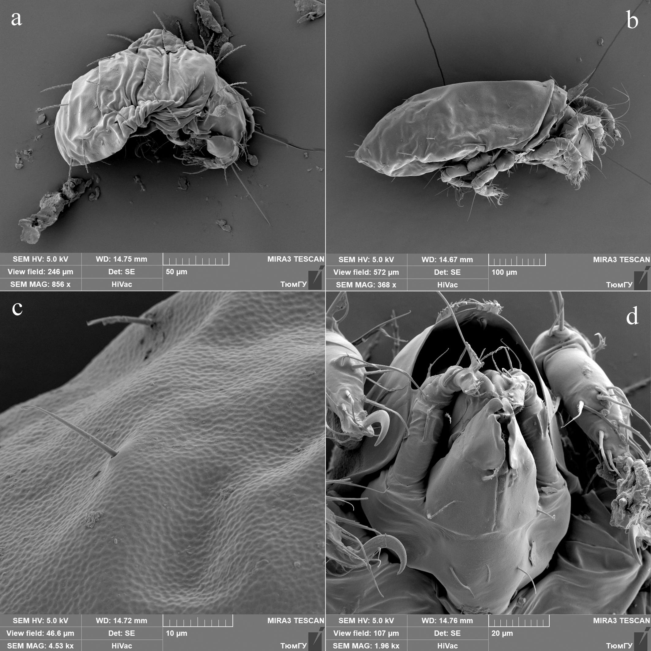
For additional understanding of adult morphology of F. spitsbergensis, we provide SEM micrographs of specimens from the Taimyr population herein (Figures 1, 2), with the ventral carinae of trochanters III and IV and femur II evident in Figures 1b, c; 2a.
Juvenile instars — Measurements – Body length of LA 210–255, PN 293–315, DN 315–390, TN 405–465. Maximum body width of LA 120–135, PN 150–165, DN 180–195, TN 225–270.
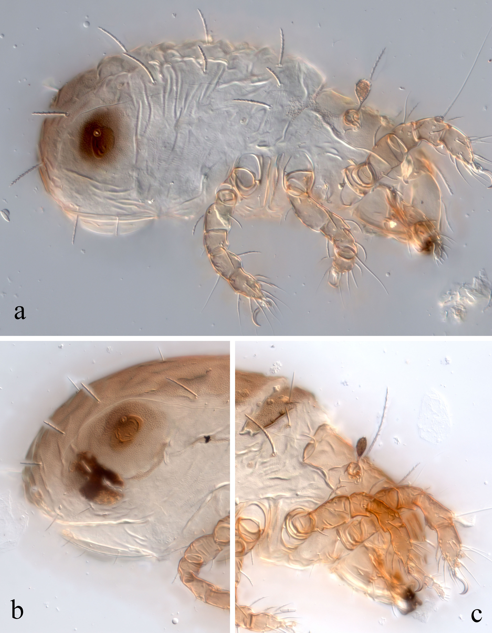
Integument (Figures 6a–c; 7a–c) – In LA, body pale, with light brown prodorsal and gastronotal shields (macrosclerites), subcapitulum, and legs. In PN, DN, and TN, body light brown, with brown prodorsal and gastronotal shields (macrosclerites), subcapitulum, and legs. Posterolateral sclerite around opisthonotal gland opening brown or dark brown. Cuticle densely microfoveolate (Fig. 6c). Lateral part of body and anogenital region sparsely plicate. Additionally, gastronotal region with transverse plications in LA (Fig. 6a).
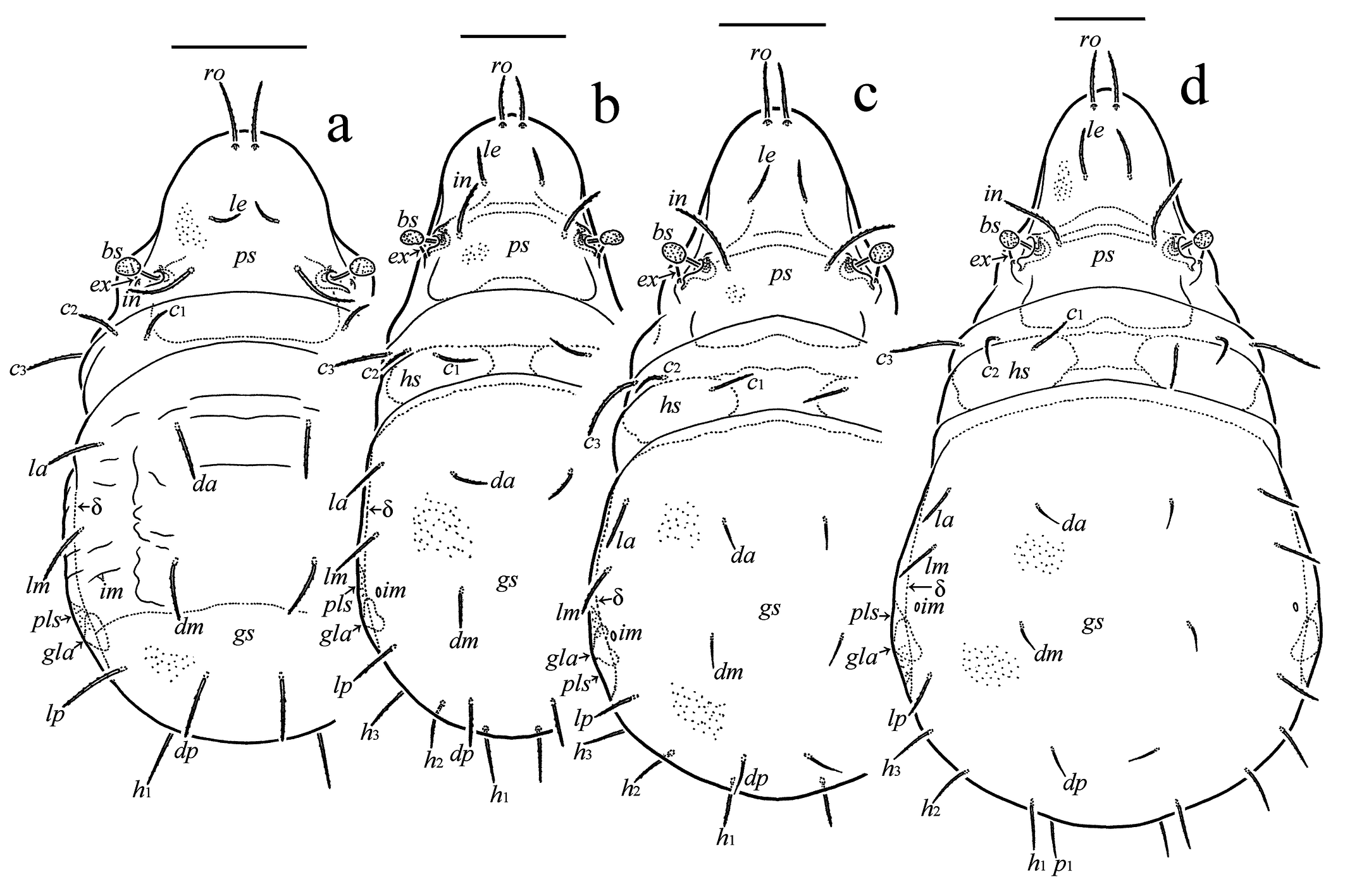
Prodorsum (Figures 3a–d; 5a, b; 6a, b; 7a, c) – Relatively short, about 1/2 (in LA) and 2/5 (in PN, DN, and TN) length of gastronotal region. Rostrum broadly rounded. Prodorsal shield (macrosclerite) well defined. Rostral (LA 30–34, PN, DN 34–37, TN 41–45), lamellar (LA 17–22, PN 22–24, DN 28–30, TN 32–37), and interlamellar (LA 37–41, PN 45–49, DN 56–64, TN 64–79) setae setiform, barbed. Bothridial seta (LA 22–26, PN, DN 26–30, TN 30–34) clavate, head rounded distally, barbed. Exobothridial seta (LA 7, PN 9, DN 13–15, TN 15) acicular. Posterior wall of bothridium forming small scale.
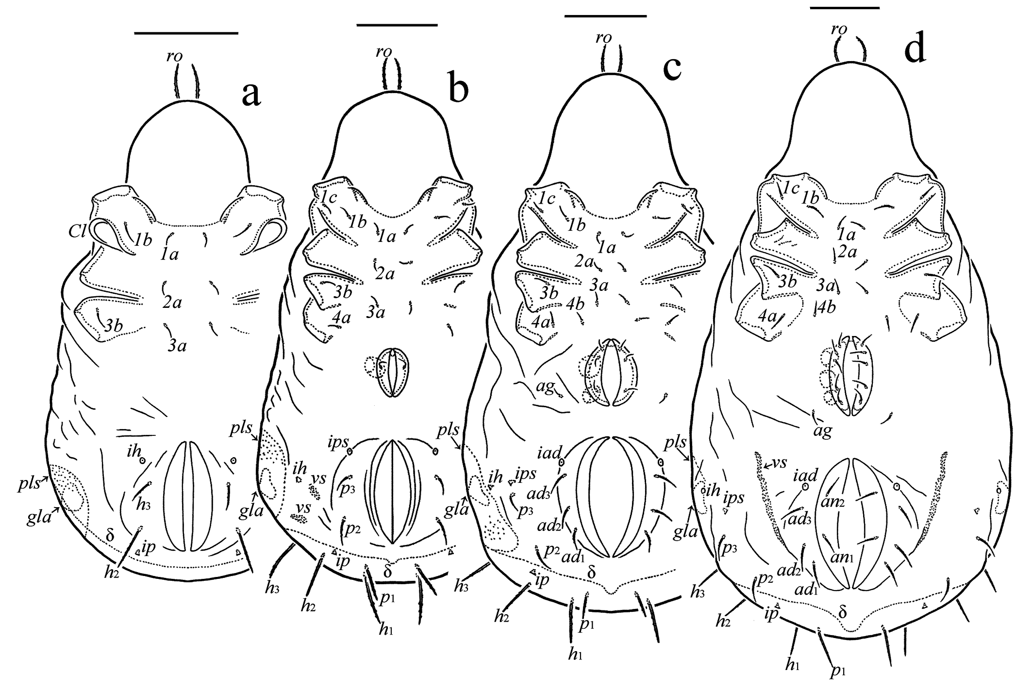
Gastronotal region (Figures 3a–d; 4a–d; 5a, b; 6a–c; 7a–c) – In LA, humeral organ absent. In PN, DN, and TN, humeral organ present, but weakly defined, positioned anterior to seta c3. In LA, gastronotal shield (macrosclerite), or pygidial sclerite, occupying posterior part of gastronotum, with anterior margin posterior to setae dm. In PN, DN, and TN, gastronotal shield occupying dorsal and posterior parts of gastronotum, with anterior margin posterior to setae of c-row. In LA, humeral sclerite absent. In PN, DN, and TN, humeral sclerite comparatively large, elongate oval, transversely oriented. In LA, lateral side of body without sclerites. In PN, DN, and TN, lateral side of body with three elongate sclerites, two of them usually fused in PN. LA with 12 pairs of setae, with dp, lp, and h1 located on gastronotal macrosclerite. PN, DN, and TN with 15 pairs of setae, with c1 located on humeral sclerite, da, la, dm, lm, dp, lp, h1–h3, and p1 located on gastronotal macrosclerite. Gastronotal setae in LA (c3, h1 26–30, c1, da, la, dm, lm, dp, lp, h2 22–26, c2 17–19, h3 13), PN (c3, h1, h3, 26–30, c1, la, lm, lp, h2 22–26, p1 19–22, c2, dm, dp 19, p2 17–19, da 15, p3 13–15), DN (c3 37, h1 26–30, c1, la, lm, lp, h2, h3 22–26, c2, p1 19–22, p2 17–19, p3 13–15, da, dm, dp 15), and TN (c3 49–52, h1 30–34, c1, la, lm, lp, h2, h3, p1 26–30, c2 22–26, p2, p3 19–22, da, dm, dp 15) setiform, barbed (except nearly smooth da, dm, and dp in DN and TN). In DN and TN, dorsocentral setae da, dm, and dp distinctly thinner than lateral setae la, lm, and lp. Sclerite absent at the base of all gastronotal setae. Two pairs of small, vaguely defined sclerites present lateral to p-segment in PN. Opisthonotal gland opening and all cupules distinct, ih and ips appearing in normal ontogenetic pattern.
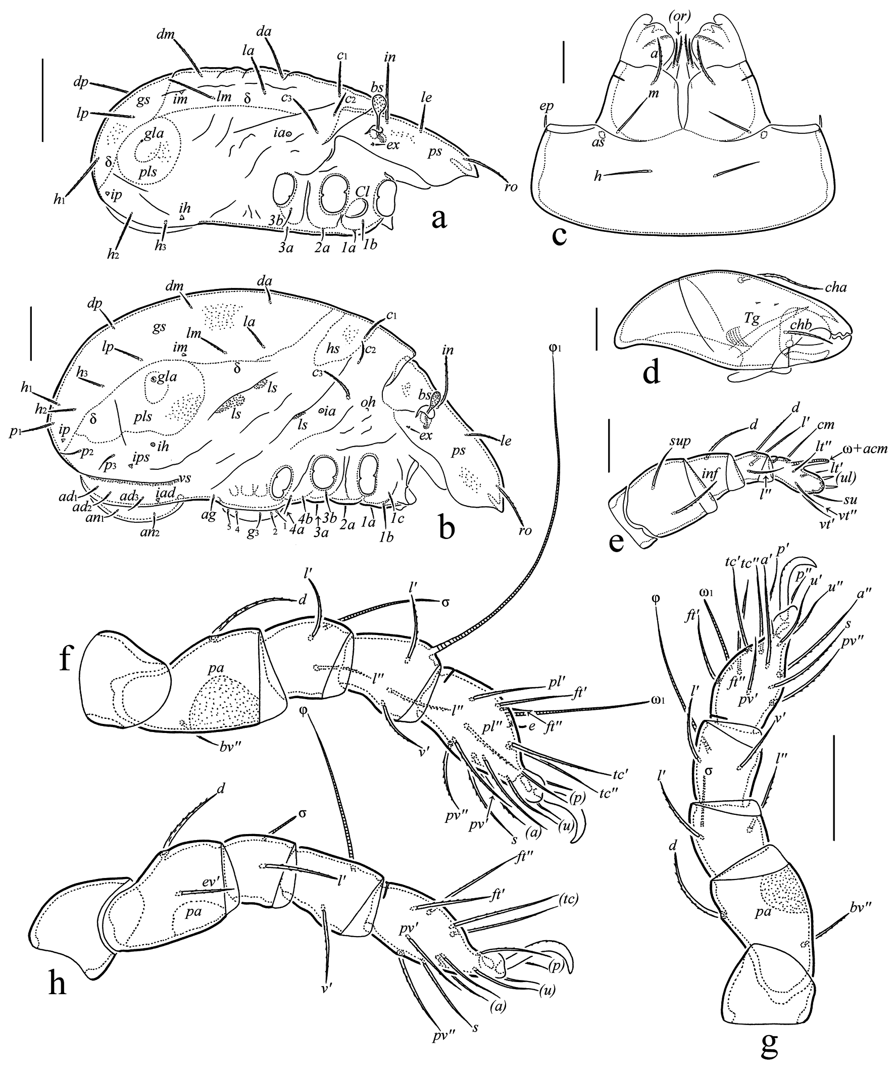
Gnathosoma (Figures 5c–e; 6d; 7a) – Subcapitulum size of LA 56 × 52, PN 64 × 60, DN 71–75 × 67–71, TN 79–82 × 75–79. Three pairs of setiform subcapitular setae a, m, and h (LA 11, PN 13, DN 15, TN 19), a barbed, m nearly smooth in LA, roughened in PN, and barbed in DN and TN, h nearly smooth. Two pairs of setiform, barbed adoral setae (LA 5, PN 7, DN 9, TN 11) . Palp length of LA 45–49, PN 49–53, DN 60, TN 67–71. Palp setal formula in all instars 0-2-1-3-9(+ω). Palpal solenidion connected with eupathidium. Tarsal seta su setiform in LA, spiniform in PN, DN, and TN. Length of postpalpal seta of LA, PN 4, DN, TN 6. Axillary saccule small, slightly oblong. Chelicera length of LA 60–64, PN 67–71, DN 82–86, TN 94–97. Cheliceral setae setiform, barbed (cha of LA 22, PN 22–24, DN 26, TN 28–30; chb of LA 15, PN 15–17, DN 19, TN 19–22).
Epimeral region (Figures 4a–d; 5a, b) – Epimeral setal formula (for successive epimeres) of LA 3-1-2 (1c as typical scale covering Claparède's organ), PN 3-1-2-1; DN and TN 3-1-2-2. Epimeral setae in LA (1a, 2a, 3a 7–9, 1b, 3b 11), PN (1a, 1c, 2a, 3a 7–9, 1b, 3b, 4a 11), DN (1a, 1c, 2a, 3a 11, 1b, 3b, 4a, 4b 13–15), and TN (1a, 1c, 2a, 3a 15, 1b, 3b, 4a, 4b 19–22) setiform, roughened.
Anogenital region (Figures 4a–d; 5a, b; 7a, b) – Ontogeny of genital (PN, DN 11, TN 13–15), aggenital (DN 11, TN 13–15), anal (TN 13–15), and adanal (DN 13–15, TN 19) setae, from LA to TN: 0-1-3-5, 0-0-1-1, 0-0-0-2, 0-0-3-3, respectively. All setae setiform, nearly smooth in LA and PN versus setiform, roughened in DN and TN. Paraproctal setae absent. Adanal cupule appearing in normal ontogenetic pattern. One pair of band-like sclerites present lateral to ad-segment in TN.
Legs (Figures 5f–h; 7a, c) – Claw of each leg strong, slightly barbed on dorsal side. Ventroparaxial porose area on femora I–IV well visible. Formulas of leg setation and solenidia of LA: I (0-2-2-3-16) [1-1-1], II (0-2-2-2-13) [1-1-1], III (0-2-1-1-13) [1-1-0]; PN: I (0-2-2-3-16) [1-1-2], II (0-2-2-2-13) [1-1-1], III (0-2-1-1-13) [1-1-0], IV (0-0-0-0-7) [0-0-0]; DN: I (0-4-2-3-16) [1-2-2], II (0-4-2-3-13) [1-1-2], III (1-2-1-2-13) [1-1-0], IV (0-2-1-1-12) [0-1-0]; TN: I (1-5-2-4-18) [1-2-2], II (1-5-2-4-15) [1-1-2], III (2-2-1-3-15) [1-1-0], IV (1-2-2-3-12) [0-1-0]. Homologies of leg setae and solenidia as indicated in Table 1.
Download as Note: Tr, Fe, Ge, Ti, Ta = trochanter, femur, genu, tibia, and tarsus, respectively. Roman letters refer to normal setae; Greek letters refer to solenidia; single prime (‘) marks setae on the anterior and double prime (’’) setae on the posterior side of a given leg segment; parentheses refer to a pair of setae. Setae are listed only for the instar in which they first appear.* ** – Seta l’’ of femora I, II appears sometimes in TN, seta v’’ of tibia IV appears sometimes in AD.
Tr
Fe
Ge
Ti
Ta
Leg I
LA
–
d, bv’’
(l), σ
(l), v’, φ1
(ft), (tc), (p), (u), (a), s, (pv), (pl), e, ω1
PN
–
–
–
–
ω2
DN
–
*(l)**
–
φ2
–
TN
v’
–
–
v’’
(it)
AD
–
v’’
–
–
–
Leg II
LA
–
d, bv’’
(l), σ
l’, v’, φ
(ft), (tc), (p), (u), (a), s, (pv), ω1
PN
–
–
–
–
–
DN
–
*(l)**
–
l’’
ω2
TN
v’
–
–
v’’
(it)
AD
–
v’’
–
–
–
Leg III
LA
–
d, ev’
l’, σ
v’, φ
(ft), (tc), (p), (u), (a), s, (pv)*
PN
–
–
–
–
–
DN
v’
–
–
l’
–
TN
l’
–
–
v’’
(it)
AD
–
–
–
–
–
Leg IV
PN
–
–
–
–
ft’’, (p), (u), (pv)
DN
–
d,* ev’*
d
v’, φ
(tc), (a), s
TN
v’
–
l’
l’,* v’’**
–
AD
–
–
–
–
–
Remarks — Main juvenile characteristics of F. spitsbergensis are as follows: bothridial seta short, clavate; humeral organ absent in LA versus weakly defined in PN, DN, and TN; gastronotal macrosclerite occupying only posterior part of gastronotum, bearing gastronotal setae dp, lp, and h1 in LA versus occupying dorsal and posterior parts of gastronotum in PN, DN, and TN; humeral sclerite absent in LA versus present, bearing gastronotal seta c1 in PN, DN, and TN; dorsocentral gastronotal setae medium-sized, barbed in LA versus progressively shorter, thinner, and with fewer barbs in PN, DN, and TN; lateral and posterolateral gastronotal setae comparatively short in PN, DN, and TN; sclerite absent at base of all gastronotal setae. Distinctive juvenile characters of F. spitsbergensis with those of other species of the genus can be found in the identification keys below.
Morphological differences of juvenile instars of F. spitsbergensis from Taimyr Peninsula (present paper) and those from Svalbard (see Table 1 in Seniczak and Seniczak 2020) are few and mainly relate to:
- Relative length of rostral and lamellar setae, with ro (34–37) slightly longer than le (28–30) in DN and TN from the Taimyr Peninsula versus ro (30) and le (31) similar in length in DN, and ro shorter than le in TN from Svalbard (despite ro shown as longer than le in Figure 10b of Seniczak and Seniczak 2020).
- Length of bothridial seta, gradually longer in each subsequent instar (LA 22–26, PN, DN 26–30, TN 30–34) in specimens from the Taimyr Peninsula versus similar in length in all instars (LA 25, PN 25, DN 25, TN 26) from Svalbard.
- Relative length of gastronotal seta h3 in LA, being 13 in LA from the Taimyr Peninsula versus 4 in LA from Svalbard (despite h3 shown as longer than 4, and longer than h2, in Figures 9a and 10a of Seniczak and Seniczak 2020).
- Relative length of gastronotal seta p3 in PN, being 13–15 in PN from the Taimyr Peninsula versus 4 in PN from Svalbard (despite p3 shown as longer than 4 in Figure 9b of Seniczak and Seniczak 2020).
- Relative length of some gastronotal setae in DN, with c3 shorter (37), da and dp similar (15), and h2–h3 similar in length (22–26) in DN from the Taimyr Peninsula versus c3 longer (49), da (10) shorter than dp (20), and h2 (20) shorter than h3 (25) in DN from Svalbard.
- Relative length of some gastronotal setae in TN, with da and dp similar in length (15), c2 (22–26), la (26–30), lp (26–30), h2 (26–30), h3 (26–30), and p3 (19–22) longer in TN from the Taimyr Peninsula versus da (10) shorter than dp (20), c2 (16), la (20), lp (18), h2 (18), h3 (22), and p3 (12) shorter in TN from Svalbard (see Table 1 in Seniczak and Seniczak 2020; however, their Figures 10b and 13 [erroneously labeled as 10 on p. 692] contradict these data, e.g., with da and dp similar in length).
Seniczak and Seniczak (2020) based their description on specimens collected by Stephen J. Coulson from Negerdalen, Edgeøya, Svalbard on 15 July 2010. In their diagnosis of F. coulsoni (p. 681), nothing contradicts the descriptions of F. spitsbergensis by Thor (1934) and Behan-Pelletier (1985), except for the presence of setae v′ on genua I, II. We consider this character variable (see below).
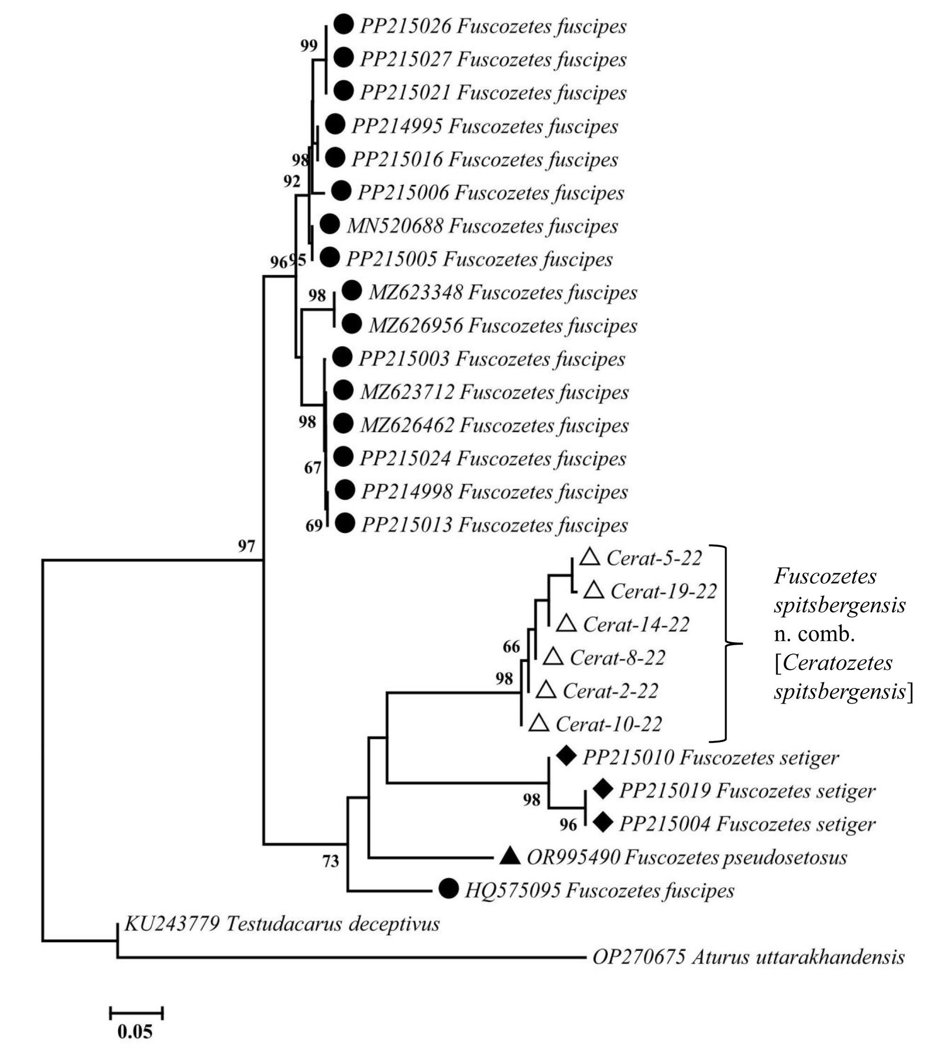
Mitochondrial genetic data — Among 19 processed individuals of F. spitsbergensis, six haplotypes were identified (Fig. 8). The resulting tree illustrates the location of them within the variability range of the genus Fuscozetes. All representatives of the genus emerged as a single branch, and all four species of the genus clustered separately with the exception of one case: HQ575095_Fuscozetes fuscipes voucher BIOUG> CAN\textgreater:DPMIT-25-32 from Canada. This case can be considered a technical error, as it was not registered by a specific author, but by the International Barcode of Life (iBOL) Consortium, and was not published. Mite haplotypes from Taimyr formed a single cluster (Fig. 8) with intra-group variability of less than 5%, confirming their single species status. The closest species to F. spitsbergensis is F. setiger, but separated by about 15% uncorrected p-distance.
Discussion
Population differences
There is small variation in genua I and II and tarsus I leg setation between adults of F. spitsbergensis by Behan-Pelletier (1985) (as C. spitsbergensis), Seniczak and Seniczak (2020) (as F. coulsoni) and herein (Table 2). We recognize that these differences possibly reflect the general population variation in specimens from different northern localities, as has been noted by Seniczak et al. (2024) for F. fuscipes, and by Behan-Pelletier (1985) for populations of some Ceratozetes species. In general, within Fuscozetes, the ontogenetically later setae, v′ on genua I, II and l″, v′ on tarsus I, can appear in tritonymph/adult stage or not (Seniczak et al. 2016, 2024; Seniczak and Seniczak 2022).
Download as Note: * − The same in specimens from Greenland (Makarova and Behan-Pelletier 2015) and Vaigach Island, Barents Sea (new data).
Segment
F. spitsbergensis (according to Behan-Pelletier 1985)
F. spitsbergensis as F. coulsoni (according to Seniczak and Seniczak 2020)
F. spitsbergensis (present data)
Genu I
v’ absent
v’ present*
v’ absent
Genu II
v’ absent
v’ present*
v’ absent
Tarsus I
l’’ and v’ present
l’’ and v’ absent
l’’ and v’ absent
Tutorium length
~113; almost reaching insertion of seta ro
107–118; almost reaching insertion of seta ro
101–116; reaching or almost reaching insertion of seta ro
After analyzing a large collection of F. spitsbergensis from different arctic regions, we attribute the differences noted between the Greenland specimens (Makarova and Behan-Pelletier 2015) and the redescription by Behan-Pelletier (1985) to normal variability. The Mongolian designation of this species as Ceratozetes cf. spitsbergensis (see Bayartogtokh 2010) is also associated with minor differences, which the author himself also considered to be a manifestation of variability (pers. com. B. Bayartogtokh, 5 January 2026).
Fuscozetes spitsbergensis–F. setiger relations
These species are undoubtedly very closely related. The diagnostic character states used in the description of adult F. setiger, F. spitsbergensis, and F. coulsoni are similar (Trägårdh 1910; Thor 1934; Behan-Pelletier 1985; Seniczak and Seniczak 2022). The presence of setae v′ on genua I, II, and setae l″ and v′ on tarsus I, as well as a degree of development of very weak pubescence of notogastral setae, vary among populations. The presence of a lateral tooth on the lamellar cusp is a rare exception in both species (Trägårdh 1910, p. 496; Behan-Pelletier 1985, p. 1332; Seniczak and Seniczak 2020, p. 682) and may also be a characteristic of individual populations or specimens, but not of a species, as noted by Seniczak and Seniczak (2022, pp. 1456, 1468). Currently, it is not possible to distinguish between these species based on adult specimens. However, the juvenile instars of F. setiger described by Seniczak and Seniczak (2022) clearly differ from those described herein as F. spitsbergensis. The assignment of adult mites to either species was apparently rather arbitrary over the course of a century. High Arctic and southern highland records were attributed primarily to the ''characteristic arctic'' Ceratozetes spitsbergensis, while Scandinavian records were attributed to F. setiger originally described from Sweden (Fig. 9). Barcoding (Fig. 8) along with juvenile morphology clearly distinguishes these very similar species. Recently, a single standard barcode fragment (COI) was considered insufficient for molecular species identification of many oribatid taxa (LeCadre et al. 2024), however the use of full-length BOLD fragments (658 bp in length) can give better results.
Ceratozetes vs. Fuscozetes
These genera are members of Ceratozetidae and, as Grandjean (1954) noted, it is difficult to separate genera in this family. They are considered members of different subfamilies, Ceratozetinae and Sphaerozetinae respectively, by Shaldybina (1972), who highlighted the presence of a ventral lamella on femur II in Fuscozetes and on femora I and II in Melanozetes. Seniczak and Seniczak (2020), along with their description of F. coulsoni, outlined states of selected morphological characters in adults of 14 species of Fuscozetes, including F. coulsoni. Most variation was found in adult notogastral setation, which varied between 10 and 11 pairs, with 10–13 pairs in F. setosus (C.L. Koch). In Seniczak and Seniczak (2020) they also questioned the inclusion of F. coulsoni in Fuscozetes because of some character states of juveniles that are more usual in species of Trichoribates in the ceratozetid subfamily Trichoribatinae, such as the presence of an opisthonotal gland sclerite (also found in juveniles of some Fuscozetes species), absence of microsclerites at some hysterosomal setae (also observed in juveniles of some Fuscozetes species), and presence of a pygidial sclerite in the larva (also found in some Fuscozetes species). However, these authors overlooked some key adult features of Fuscozetes species: the bothridium with evident medial and lateral scales, rather than cup-shaped, and the lack of horizontal folds in the integument dorsad of acetabula II and III, states found in all members of Trichoribatinae.
Distributional, ecological and biological remarks
Distribution
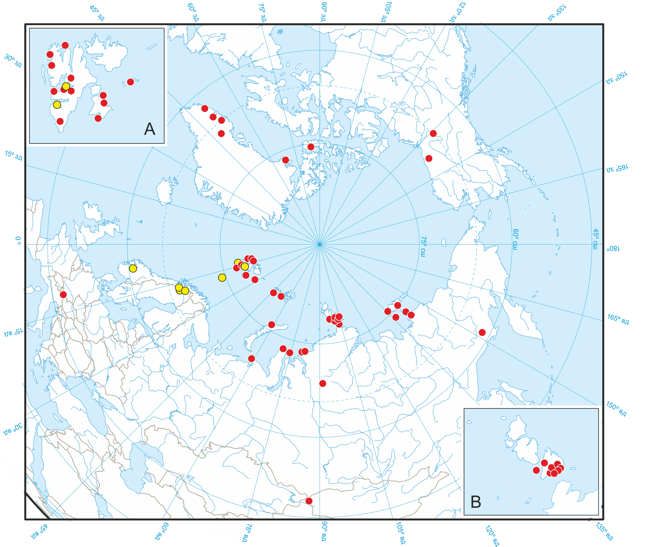
Traditionally, the distribution of many characteristic arthropod species of the Arctic plains that also occur in southern mountains, is designated as arctic-alpine (Reiss et al. 1999; Marusik and Eskov 2009; Fischer et al. 2016). In the case of soil microarthropods, the non-arctic occurrences are usually associated with the mountain systems of southern Siberia (Babenko and Fjellberg 2001; Bayartogtokh 2010; Lindquist and Makarova 2012; Makarova 2013; Makarova and Lindquist 2013; Potapov et al. 2018). Only a few ''arctic'' species inhabit Siberian and European mountains (Fjellberg 1986; Makarova et al. 2010). Fuscozetes spitsbergensis (Fig. 9) belongs to this group (Fischer et al. 2016).
Cryophily
Fuscozetes spitsbergensis is consistently found in very cold habitats (Table 3). In extreme cold polar desert regions, this species occurs at altitudes close to sea level (usually no higher than 100 m a.s.l.), while in the subarctic mountains it is found in tundra and nival-desert belts at mid-altitudes (300–1,600 m a.s.l.), and in the Austrian Alps and Mongolian Altai it is recorded only in the highlands, at altitudes of 2,800–3,300 m a.s.l. (Bayartogtokh 2010; Fischer et al. 2016). In the Tyrolean mountains, where soil mites were studied along an altitudinal gradient of 2,700–3,400 m a.s.l., F. spitsbergensis was found only at 3,300 m a.s.l., where average monthly temperatures measured at a depth of 10 cm ranged between -10.3 °C and +2.9 °C (Fischer et al. 2016). On the Putorana Plateau, southern Taimyr, where soil microarthropods were studied along an altitudinal profile in the range of 350–900 m a.s.l. (from the shrub tundra belt to nival deserts), F. spitsbergensis was found only in the damp nival deserts on the 700 m a.s.l. terrace (single specimen) and on the 900 m a.s.l. terrace (numerous specimens, Table 4). It was reasonably assumed that the average July temperature on the 900 m a.s.l. terrace does not exceed 2−3 °C (Matveyeva 2002).
Download as
Latitudinal position
Country, region
Locality
Latitude
Minimal altitude, m a.s.l.
Biotope
Source
HIGH ARCTIC
Russia, Franz Josef Land, Hooker Island
Tikhaya Bay
80°20′N
10−15
Slope sparse desert-like community dominated by Luzula confusa, Alopecurus alpinus, Aulacomnium turgidum, Racomitrium sp., Stereocaulon alpinum; tundra-like well drained community with Salix polaris, Alopecurus alpinus, Papaver polare, Cerastium arcticum, Saxifraga spp., Racomitrium sp., Aulacomnium turgidum, lichens
Makarova 2023 [as Ceratozetes spitsbergensis], with A. Babenko’s (2026) clarification
Russia, Severnaya Zemlya Archipelago, Bolshevik Island
Solnechnaya Bay
78°12′N
10−15
Kurum with Racomitrium lanuginosum and lichens; tundra-like, well drained community with Saxifraga cespitosa, Papaver polare, Salix polaris, mosses; polygonal community on sandy grounds dominated by Gymnomitrion corallioides and Racomitrium lanuginosum; lemming hills with Alopecurus alpinus, Poa spp., Papaver polare, Luzula spp., mosses
Makarova 2002a [as Ceratozetes spitsbergensis]
Russia, N Taimyr
Chelyuskin Peninsula
77°41′N
3−5
Kurum with cushions of moss Racomitrium lanuginosum
New data
Norway, Svalbard, Edgeøya
Negerdalen
77°18′N
?
Data not available
Ávila‑Jiménez et al. 2019 [as Ceratozetes spitsbergensis]
4
Sparse graminoid vegetation with Luzula sp., moss Racomitrium lanuginosum, lichens
Seniczak and Seniczak 2020 [as Fuscozetes coulsoni]
Russia, W Taimyr
Dikson Island
73°31′N
35−40
Zonal tundra dominated by Salix polaris, Carex arctisibirica, Luzula sp, mosses, lichens
New data
Russia, West Siberia, Kara Sea
Shokalsky Island
72°58′N
7−10
Dryad tundra on hill slope with Salix nummularia, Dryas punctata, Vaccinium vitis-idea, Alopecurus alpinus, mosses, lichens; zonal tundra on well drained watershed, with Carex arctisibirica, Poa alpigena, Luzula confusa, mosses, lichens
Bizin and Makarova 2024 [as Ceratozetes spitsbergensis]
SUBARCTIC
Russia, southern Taimyr, Putorana Plateau
Yt-Kyuel (Sobachie) Lake vicinity
69°01′N
700
Wet nival desert on upper plateau terraces with Deschampsia borealis, Cerastium regelii, Raninculus sulphureus, Saxifraga spp., Cardamine bellidifolia, Dicranoweisia crispula, Racomitrium canescens. Cladonia spp. Cetrariella delisei
Matveyeva 2002 [vegetation]; new data
USA, Alaska, Brooks Range
“Budweiser Mountain”
68°12′N
1200
Alpine tundra, mosses
Behan-Pelletier 1985 [as Ceratozetes spitsbergensis]
Denmark, SW Greenland
Eqaluit, Bjørnesund
62°54′N
300
Snow bed with willow, mosses, lichens
Makarova and Behan-Pelletier 2015, with A. Fjellberg’s (2017) clarification [as Ceratozetes sp. near spitsbergensis]
Russia, Far East, Magadan Region
Aborigen Pik vicinity
61°58′N
1400
Snow bed run-off, wet vegetation with Phyllodoce sp. and mosses; crevices on rocks, rather dry moss and lichen
Behan-Pelletier 1985 [as Ceratozetes spitsbergensis]
SOUTH HIGHLANDS
Mongolia, Altai Tavan Bogd
Bayan-Ölgii Aimak
48°36′N
2800
Wet alpine meadows with mosses
Bayartogtokh 2010 [as Ceratozetes cf.spitsbergensis] with clarification (Fisher et al. 2016)
Austria, Tyrol, Stubai Alps
Schrankogel Mt.
47°03′N
3300
Alpine-nival cushion plant community dominated by Androsace alpina, Poa laxa, Saxifraga bryoides, Cerastium uniflorum, Ranunculus glacialis, Polytrichum piliferum, Thamnolia vermicularis
Fisher et al. 2016 [as Ceratozetes spitsbergensis]
Abundance and occurrence
The highest abundance of F. spitsbergensis (140–1200 ind./dm2) was recorded precisely at the limit of heat supply in the polar desert zone (Franz Josef Land, Severnaya Zemlya Archipelago, Chelyuskin Peninsula on Taimyr), where the average July temperature varies between +1.2 and +2.2 °C (Table 4). These mites are found only in habitats with full drainage (kurums, dwarf willow-forb-mossy communities on slopes, lemming hills, etc.), but avoid the eutrophication of bird cliff colonies and lemming burrows (Makarova 2002a, 2023). Their occurrence (in samples with an area of 25 cm²) in such drained habitats is generally high, 76–100% (recalculated from data used in Makarova 2002a, 2023). In the tundra on Edgeøya Island, Svalbard, F. spitsbergensis was found in only two of 118 soil samples (Seniczak and Seniczak 2020). The same is true for the nival belt of the central Eastern Alps, where almost all individuals (384 from 388) were found in only one of 56 samples of 20.3 cm2 each (Fischer et al. 2016).
Download as Note: * − Total 2286 individuals; ** − from Makarova 2002a.
Nature zone/subzone/belt
Region, collector, date
Latitude
Altitude, m a.s.l.
Biotope/habitat (number of samples 5x5 cm)
Number of studied individuals*
Abundance, ind./dm2
Part of males among adults, %
Part of gravid females, %
Part of juveniles, %
Polar desert
Franz Josef Land, Hooker Island, Tikhaya Bay, 14.07.2015, A. Babenko
80°20′N
10−15
Dwarf willow-forb-mossy communities (8)
847
424
57
72
30
Polar desert
Severnaya Zemlya, Krasnoflotskie Ostrova, 30.08.2019, M.V. Gavrilo
78°34′N
≈ 20
Nest of Larus heuglini (2)
158
316
50
80
66
Polar desert
Severnaya Zemlya, Bolshevik Island, Solnechnaya Bay, 26.07.2000, O. Makarova (I)
78°12′N
10−15
Kurum covered by mosses and lichens (10)
424
1190**
54
53
57
Ibid., 05.08.2000, O. Makarova (II)
Dwarf willow-forb-mossy communities (10)
341
210**
46
59
83
Polar desert
N Taimyr, Chelyuskin Peninsula, 09.08.2025, P. Pavlova
77°41′N
3−5
Kurum covered by Racomitrium lanuginosum (7)
244
139
43
25
61
Arctic tundra
Novaya Zemlya, North Island, Arkhangelskaya Bay, 29.08.1996, G. Khakhin
75°52′N
20−35
Lichen tundra (4)
98
98
51
11
24
Arctic tundra
W Taimyr, Dikson Island, 02.09.2000, O. Makarova
73°31′N
35−40
Dwarf willow-forb-mossy tundra (10)
72
29
59
0
69
Nival desert
S Taimyr, Putorana Plateau, Yt-Kyuel Lake, 27.07.1996, O. Makarova
69°01′N
900
Wet forb-lichen-mossy nival desert (10)
102
41
31
84
29
Demography
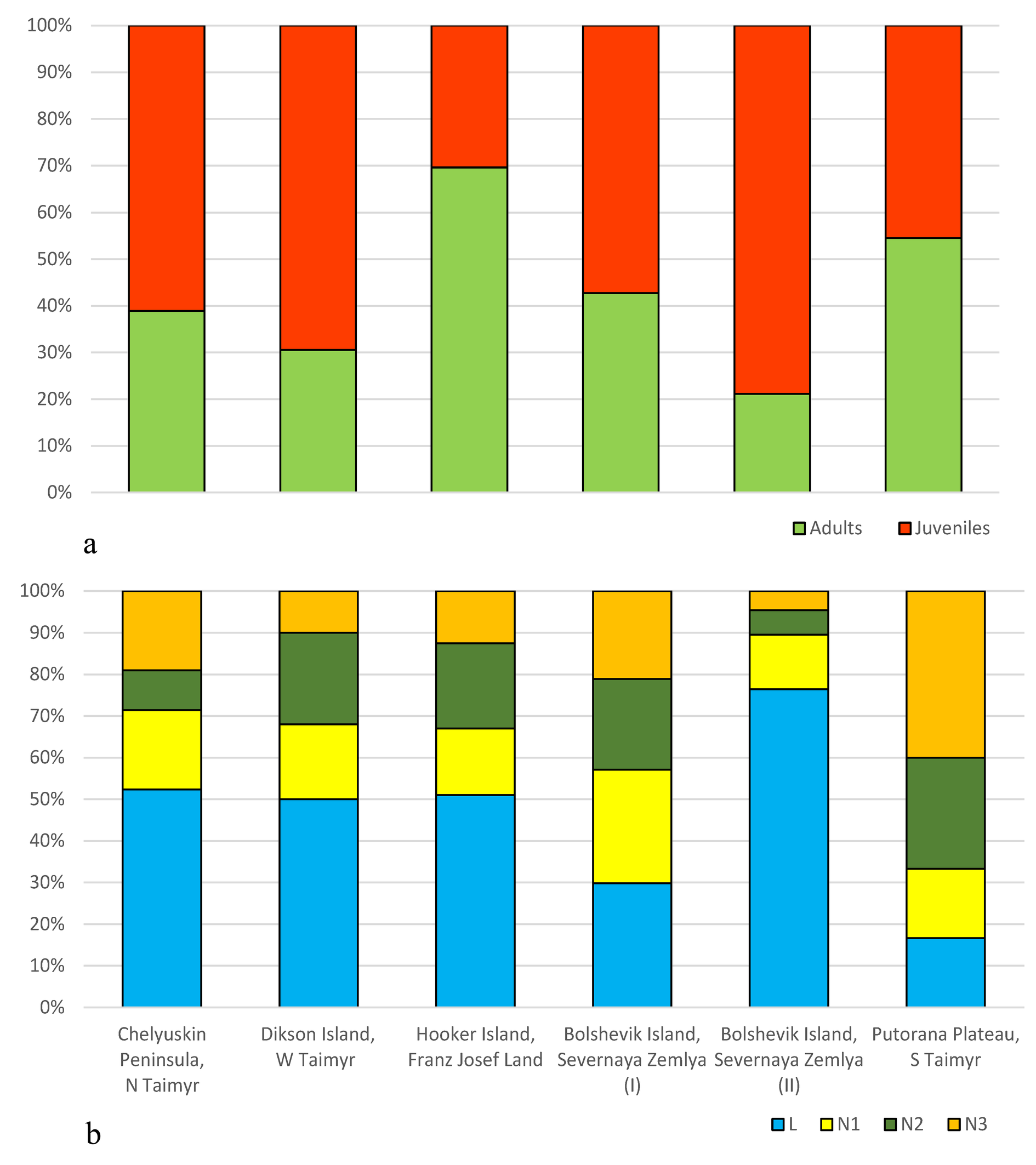
We analyzed the summer demographic structure of F. spitsbergensis in seven regions of the northern Palearctic, using 2,286 specimens (Table 4). The proportion of males in samplings varied widely (31–59%), as did the proportion of gravid females at the height of summer (43–84%). At the end of August, the number of females with eggs decreased (11%), and in September, females lacked eggs. One, two, three, or four eggs can develop simultaneously in one female (41%, 39%, 17%, and 3% of cases, respectively). All active ontogenetic instars were present in all samplings, indirectly indicating multi-year development. The fraction of juvenile individuals in some samplings constituted 24–83% of the population (Fig. 10a), with a sharp predominance of larvae (50–77%) sometimes observed, even in September (Fig. 10b). In different biotopes within one locality (Bolshevik Island, Solnechnaya Bay, I and II), the demographic structure of the population varied greatly (Fig. 10a, b).
Feeding
In the laboratory, all stages of F. spitsbergensis fed on dead springtails Folsomia binoculata (Wahlgren, 1899), Isotomidae, and various species of lichens, including Sphaerophorus globosus, Sphaerophorus fragilis, Parmelia omphalodes, and Megaspora verrucosa (Makarova 2002b, new data).
Key to larvae of Fuscozetes
1. Gastronotal shield (macrosclerite, pygidial sclerite) absent
...... 2
— Gastronotal shield (macrosclerite, pygidial sclerite) present
...... 3
2. Humeral sclerite absent. Bothridial seta noticeably shorter than interlamellar seta, clavate. Gastronotum without additional sclerites. Centrodorsal gastronotal setae short, smooth. All gastronotal setae without microsclerite at their base. Humeral organ absent
...... F. kamchatkicus Seniczak, Kaczmarek and Seniczak, 2016 (see Seniczak et al. 2016).
— Humeral sclerite present. Bothridial seta longer than interlamellar seta, narrowly fusiform. Gastronotum with several additional large and numerous small sclerites. Centrodorsal gastronotal setae medium-sized, barbed. Some gastronotal setae with microsclerite at their base. Humeral organ present
...... Two similar species, F. fuscipes (C.L. Koch, 1844) and F. pseudosetosus (Shaldybina, 1975) (see Shaldybina 1969, 1977, 1978; Seniczak 1989; Seniczak et al. 2024)
3. Gastronotal shield (pygidial sclerite) occupying only pygidial part of gastronotum. Humeral sclerite absent. Bothridial seta noticeably shorter than interlamellar seta
...... 4
— Gastronotal shield (pygidial sclerite) occupying dorsal and pygidial parts of gastronotum. Humeral sclerite present. Bothridial seta longer than interlamellar seta or nearly equal in length
...... 5
4. Humeral organ absent
...... F. spitsbergensis (Thor, 1934) n. comb. (= F. coulsoni Seniczak and Seniczak, 2020 n. syn.) (see Seniczak et al. 2020; present data).
— Humeral organ present
...... F. setiger (Trägårdh, 1910) (see Seniczak and Seniczak 2022)
5. Gastronotum medially with several additional small sclerites, located in transverse row. Gastronotal seta c1 anteromedial of humeral sclerite. Bothridial seta broadly fusiform
...... F. tatricus Seniczak, 1993 (see Seniczak 1993)
— Gastronotum without additional sclerites medially. Gastronotal seta c1 associated with humeral sclerite. Bothridial seta narrowly fusiform
...... F. setosus (C.L. Koch, 1839) (see Seniczak 1989)
Key to tritonymphs of Fuscozetes
Nymphal instars are similar in many morphological characteristics, therefore, this key can also be applied to protonymphs and deutonymphs (except characteristics that are specific to each instar as, for example, body size and number of genital setae).
1. Gastronotal seta c1 distanced from humeral sclerite
...... 2
— Gastronotal seta c1 associated with humeral sclerite
...... 4
2. Bothridial seta clavate. Gastronotal setae of c series with microsclerite at their base. Other gastronotal setae on macrosclerite. Lamellar seta barbed
...... F. tatricus Seniczak, 1993 (see Seniczak 1993)
— Bothridial seta narrowly fusiform. Some gastronotal setae with microsclerite at their base. Lamellar seta smooth
...... 3
3. Dorsocentral (da, dm) and posterolateral (h1, h2) gastronotal setae slightly different in length
...... Two similar species, F. fuscipes (C.L. Koch, 1844) and F. pseudosetosus (Shaldybina, 1975) (see Shaldybina 1969, 1977, 1978; Seniczak 1989; Seniczak et al. 2024)
— Posterolateral (h1, h2) gastronotal setae longer than dorsocentral gastronotal setae (da, dm)
...... F. setosus (C.L. Koch, 1839) (see Seniczak 1989)
4. Gastronotal seta h1 noticeably shorter than half of distance dp–h1
...... F. spitsbergensis n. comb. (Thor, 1934) (= F. coulsoni Seniczak and Seniczak, 2020 n. syn.) (see Seniczak et al. 2020; present data)
— Gastronotal seta h1 longer than half of distance dp–h1
...... 5
5. Humeral organ absent
...... F. kamchatkicus Seniczak, Kaczmarek and Seniczak, 2016 (see Seniczak et al. 2016)
— Humeral organ present
...... F. setiger (Trägårdh, 1910) (see Seniczak and Seniczak 2022)
Acknowledgements
Our special thanks to V. Kirnev (University of Tyumen, Tyumen, Russia) for SEM micrographs, and two anonymous reviewers for their valuable comments. The sampling on the Taimyr by P.D. Pavlova and I.A. Smirnov was the part of the scientific program of ''Great Arctic Expedition 2025'' organized by the Moscow Centre of Supplemental Education ''Travel Laboratory''. Arrangement of the map was prepared for publication with helpful participation of K.V. Makarov (Moscow Pedagogical State University, Russia). This study was supported by the Ministry of Science and Higher Education of the Russian Federation within the framework of the Carbon Measurement Test Area in Tyumen′ Region (FEWZ-2024-0016). Besides, we are greatly obliged to A. Fjellberg (Tjøme, Norway), B. Bayartogtokh (National University of Mongolia), A.B. Babenko, V.I. Bulavintsev (Severtsov Institute of Ecology and Evolution, Moscow, Russia), N.V. Matveyeva (Komarov Botanical Institute, St. Petersburg, Russia), and M.V. Gavrilo (Arctic and Antarctic Research Institute, St. Petersburg, Russia) for the useful consultations and/or material, as well as to R.A. Norton (New York-College of Environmental Science and Forestry in Syracuse, USA) for use of his wonderful library.
References
- Ávila-Jiménez M.L., Solhøy, T., Gwiazdowicz D.J., Fjellberg A., Dózsa-Farkas K., Monson F., De Smet W.H., Stur E., Ekrem T., Coulson S.J. 2019. The terrestrial invertebrate fauna of Edgeøya, Svalbard: Arctic landscape community composition reflects biogeography patterns. Polar Biol., 42, 837-850 . https://doi.org/10.1007/s00300-019-02471-x
- Babenko A.B., Fjellberg A. 2001. A new species of Morulina (Collembola, Neanuridae, Morulininae) from Siberia. Norw. J. Ent., 48: 275-280.
- Bayartogtokh B. 2010. Oribatid mites of Mongolia (Acari: Oribatida). Moscow: KMK Scientific Press Ltd. 371 pp. [in Russian]
- Behan-Pelletier V.M. 1985. Ceratozetidae of the western North American arctic. Can. Ent., 117: 1287-1366. https://doi.org/10.4039/Ent1171287-11
- Behan-Pelletier V.M. 1997. Oribatid mites (Acari: Oribatida) of the Yukon. In: Danks H.V., Downes J.A. (Eds.). Insects of the Yukon. Ottawa: Biological survey of Canada (Terrestrial Arthropods), Canadian Museum of Nature. pp. 115-149.
- Behan-Pelletier V.M., Eamer B. 2009. Ceratozetes and Ceratozetoides (Acari: Oribatida: Ceratozetidae) of North America. Can. Ent., 141: 246-308. https://doi.org/10.4039/n09-023
- Behan-Pelletier V., Schatz H. 2010. Patterns of diversity in the Ceratozetoidea (Acari: Oribatida): a North American assessment. In: Sabelis M.W., Bruin J. (Eds.). Trends in Acarology. Proc. XII Int. Cong. Acarology, Amsterdam (2006). pp. 97-104. https://doi.org/10.1007/978-90-481-9837-5_15
- Berlese A. 1908. Elenco di generi e specie nuovi di Acari. Redia, 5: 1-15.
- Bizin M., Makarova O.L. 2024. Free-living mites (Acari) of the Shokalsky Island, off the northern Gyda Peninsula, Kara Sea, High Arctic. Acarologia, 64: 172-191. https://doi.org/10.24349/634f-jb4i
- Chernov Yu.I. 2004.The animal world of the polar desert on the Devon Island Plateau, Canadian Arctic Archipelago. Entom. Rev., 84: 15-24.
- Coulson S.J., Leinaas H.P., Ims, R.A., Søvik G. 2000. Experimental manipulation of the winter surface ice layer: the effects on a High Arctic soil microarthropod community. Ecography, 23: 299-306. https://doi.org/10.1111/j.1600-0587.2000.tb00285.x
- Coulson S.J., Fjellberg A., Melekhina E.N., Taskaeva A.A., Lebedeva N.V., Belkina O., Seniczak S., Seniczak A., Gwiazdowicz D.J. 2015. Microarthropod communities of industrially disturbed or imported soils in the High Arctic; the abandoned coal mining town of Pyramiden, Svalbard. Biodivers. Conserv., 24: 1671-1690. https://doi.org/10.1007/s10531-015-0885-9
- Coulson S.J., Convey P., Aakra K. Aarvik L. Ávila-Jiménez M.L., Babenko A., Biersma E.M., Boström S., Brittain J.E., Carlsson A.M., Christoffersen K., De Smet W.H., Ekrem T., Fjellberg A., Füreder L. Gustafsson D. Gwiazdowicz D.J., Hansen L.O., Holmstrup M., Hullé M., Kaczmarek L. Kolicka M., Kuklin V., Lakka H.-K., Lebedeva N., Makarova O., Maraldo K., Melekhina E., Ødegaard F., Pilskog H.E., Simon J.C:, Sohlenius B., Solhoy T., Soli G., Stur E., Tanasevitch A., Taskaeva A., Velle G., Zawierucha K., Zmudczynska-Skarbek K. 2014. The terrestrial and freshwater invertebrate biodiversity of the archipelagoes of the Barents Sea; Svalbard, Franz Josef Land and Novaya Zemlya. Soil Biol. Biochem., 68: 440-470. https://doi.org/10.1016/j.soilbio.2013.10.006
- Dalenius P. 1960. Studies on the Oribatei (Acari) of the Torneträsk Territory in Swedish Lappland. I. A list of the habitats and the composition of their oribatid fauna. Oikos, 11, 80-124. https://doi.org/10.2307/3564885
- Fischer B.M., Schatz H., Querner P., Pauli H. 2016. Ceratozetes spitsbergensis Thor, 1934: an arctic mite new to Continental Europe (Acari: Oribatida). Int. J. Acarol., 42: 135-139. https://doi.org/10.1080/01647954.2015.1133702
- Fjellberg A. 1986. Revision of the genus Agrenia Borner, 1906 (Collembola: Isotomidae). Ent. scand., 17: 93-106. https://doi.org/10.1163/187631286X00134
- Folmer O., Black M., Hoeh W., Lutz R., Vrijenhoek R. 1994. DNA Primers for Amplification of Mitochondrial Cytochrome C Oxidase Subunit I from Diverse Metazoan Invertebrates. Mol. Marine Biol. Biotech., 3: 294-299.
- Grandjean F. 1954. Essai de classification des Oribates (Acariens). Bull. Soc. zool. France, 78: 421-446.
- Grandjean F. 1963. Concernant Sphaerobates gratus, les Mochlozetidae et les Ceratozetidae (Oribates). Acarologia, 5: 284-305.
- Jørgensen M. 1934. Ein revidiertes Verzeichnis űber grönländische Milben. Zool. Anz., 107: 40−47.
- Karppinen E. 1967. Notes on the Arthropod fauna of Spitzbergen. 2. Data on the oribatid (Acari) of Spitzbergen. Ann. Ent. Fenn., 33: 18-26.
- Koch C.L. 1839. Deutschlands Crustaceen, Myriapoden und Arachniden, Heft 30. F. Pustet, Regensburg.
- Koch C.L. 1844. Deutschlands Crustaceen, Myriapoden und Arachniden, Heft 38. F. Pustet, Regensburg.
- Krivolutsky D.A., Lebrun P., Kunst M., Akimov I.A., Bayartogtokh B., Vasiliu N., Golosova L.D., Grishina L.G., Karppinen E., Kramnoy V.Ya., Laskova L.M., Luxton M., Marshall V., Matveenko A.A., Netuzhilin I.A., Norton R., Sitnikova L.G., Smrž J., Starý J., Tarba Z.M., Shaldybina E.S., Еitminavichute I. 1995. Oribatid mites: Morphology, Development, Phylogeny, Ecology, Methods of Study and Characteristics of the Model Species Nothrus palustris. Moscow: Nauka Publisher. 224 pp. [In Russian]
- Kumar S., Stecher G., Tamura K. 2016. MEGA7: Molecular Evolutionary Genetics Analysis version 7.0 for bigger datasets. Mol. Biol. Evol., 33: 1870-1874. https://doi.org/10.1093/molbev/msw054
- Lebedeva N.V., Lebedev V.D., Melekhina E.N. 2006. New data on the oribatid mite (Oribatei) fauna of Svalbard. Dokl. Biol. Nauk, 407: 182-186. [in Russian] https://doi.org/10.1134/S0012496606020207
- Lebedeva N.V., Melekhina E.N., Lebedev V.D. 2014. Oribatid mites in habitats and nests of the snow bunting in the high Arctic. In: Complex Investigations of the Svalbard and Offshore Nature. Proceedings of the International Scientific Conference (Murmansk, 6-8 November 2014). Issue 12. Moscow: GEOS. pp. 162-168. [In Russian]
- Le Cadre J., Klemp F.L., Bálint M., Scheu S., Schaefer I. 2024. Applicability and perspectives for DNA barcoding of soil invertebrates. PeerJ, 12: e17709 https://doi.org/10.7717/peerj.17709
- Lindquist L.L., Makarova O.L. 2012. Review of the mite subfamily Arctoseiinae Evans with a key to its genera and description of a new genus and species from Siberia (Parasitiformes, Mesostigmata, Ascidae). ZooKeys, 233: 1-20. https://doi.org/10.3897/zookeys.233.3862
- Makarova O.L. 2002a. Acarocenoses (Acariformes, Parasitiformes) in polar deserts: 1. Mite assemblages of the Severnaya Zemlya Archipelago: structure of fauna and abundance. Ent. Rev., 82: 839-856.
- Makarova O.L. 2002b. Acarocenoses (Acariformes, Parasitiformes) in polar deserts. 2. Cenotic relations, structure of communities, and proportion of suborders. Ent. Rev., 82: 857-875.
- Makarova O.L. 2013. Gamasid mites (Parasitiformes, Mesostigmata) of the European Arctic and their distribution patterns. Ent. Rev., 93: 113-133. https://doi.org/10.1134/S0013873813010156
- Makarova O.L. 2023. Free-living mites (Acari) of the Franz Josef Land Archipelago, the coldest Old World territory: diversity, geographic distributions, assemblages. Acarologia, 63: 1163-1186. https://doi.org/10.24349/p6wb-pcni
- Makarova O.L., Behan-Pelletier V.M. 2015. Oribatida (=Cryptostigmata, Beetle mites). In: Böcher J., Kristensen N.P., Pape T., Vilhelmsen L. (Eds.). The Greenland Entomofauna. An Identification Manual of Insects, Spiders and Their Allies. Leiden - Boston: Brill. pp. 802-845.
- Makarova O.L., Lindquist E.E. 2013. A new species of the gamasid mite genus Arctoseius Thor, 1930 (Parasitiformes, Mesostigmata, Ascidae) from Russia with a key to the multidentatus species-group. ZooKeys, 313: 9-24. https://doi.org/10.3897/zookeys.313.5317
- Makarova O.L., Ermilov S.G., Yurtaev A.A., Mansurov R.I. 2015. The first data on soil mites (Acari) of Arctic Belyi Island (Northern Yamal, the Kara Sea). Zool. Zh., 94: 899-904. [in Russian] https://doi.org/10.1134/S0013873815060147
- Makarova O.L., Osadtchy A.V., Melnikov M.V. 2010. Gamasid Mites (Parasitiformes, Mesostigmata) in nests of passerine birds on the arctic Seven Islands Archipelago, the Barents Sea. Ent. Rev., 90: 643-649. https://doi.org/10.1134/S0013873810050118
- Marusik Y.M., Eskov K.Y. 2009. Spiders (Arachnida: Aranei) of the tundra zone of Russia. In: S.I. Golovatch, O.L. Makarova, A.B. Babenko, Penev L.D. (Eds). Species and Communities in Extreme Environments. Sofia: Pensoft Publishers. pp 131-164.
- Matveyeva N.V. 2002. Association Dicranoweisio-Deschampsietum ass. nov. in cold desert belt of Putorana Plateau (Srednesibirskoe Plateau). Vegetation of Russia, 3: 32-41. https://doi.org/10.31111/vegrus/2002.03.32
- Nicolet H. 1855. Histoire naturelle des Acariens qui se trouvent aux environs de Paris. Arch. Mus. Hist. Nat., 7: 381-482. https://doi.org/10.5962/bhl.title.66066
- Niedbała W. 1971. Oribatei (Acari) of Spitsbergen. B. Pol. Acad. Sci. Biol., 19: 737-742.
- Norton R.A. 1977. A review of F. Grandjean's system of leg chaetotaxy in the Oribatei (Acari) and its application to the family Damaeidae. In: Dindal D.L. (Ed.). Biology of Oribatid Mites. Syracuse: SUNY Col. Env. Sci. Forestry. pp. 33-61.
- Norton R.A., Behan-Pelletier V.M. 2009. Suborder Oribatida. Chapter 15. In: Krantz G.W., Walter D.E. (Eds). A Manual of Acarology. Lubbock: Texas Tech Univ. Press. pp. 430-564.
- Norton R.A., Ermilov S.G. 2014. Catalogue and historical overview of juvenile instars of oribatid mites (Acari: Oribatida). Zootaxa, 3833: 1-132. https://doi.org/10.11646/zootaxa.3833.1.1
- O'Neill J.C., Fisher J.R., Nelson W.A., Skvarla M.J., Fisher D.M., Dowling A.P.G. 2016. Systematics of testudacarine torrent mites (Acari, Hydrachnidia, Torrenticolidae) with descriptions of 13 new species from North America. ZooKeys, 582: 13-110. https://doi.org/10.3897/zookeys.582.7684
- Pešić V., Smit H., Rayal R., Sharma N., Rana K.K., Bahuguna P. 2022. First DNA barcodes of water mites from the Indian Himalayas with description of two new species (Acari, Hydrachnidia). Int. J. Acarol., 48: 479-485. https://doi.org/10.1080/01647954.2022.2130424
- Potapov M., Babenko A., Fjellberg A., Schulz H.-J. 2018. Taxonomy of a predaceous springtail: the revision of the Palaearctic species of Metisotoma Maynard, 1951. Zootaxa, 4399: 69-86. https://doi.org/10.11646/zootaxa.4399.1.4
- Reiss R.A., Ashworth A.C., Schwert D.P. 1999. Molecular genetic evidence for the post-Pleistocene divergence of populations of the arctic-alpine ground beetle Amara alpina (Paykull) (Coleoptera: Carabidae). J. Biogeogr., 26: 785-794. https://doi.org/10.1046/j.1365-2699.1999.00321.x
- Sellnick M. 1928. Formenkreis: Hornmilben, Oribatei. In: Brohmer P., Ehrmann P., Ulmer G. (Eds). Die Tierwelt Mitteleuropas. Leipzig: Quelle und Meyer, (3) 9. pp. 1-42.
- Seniczak A., Seniczak S. 2020. Morphological ontogeny of Fuscozetes coulsoni sp. nov. (Acari: Oribatida: Ceratozetidae) from Svalbard, Norway. Syst. Appl. Acarol., 25: 680-696. https://doi.org/10.11158/saa.25.4.8
- Seniczak A., Seniczak S. 2022. Systematic position of Fuscozetes setiger (Acari: Oribatida: Ceratozetidae) in the light of ontogenetic studies. Syst. Appl. Acarol., 27: 1454-1474. https://doi.org/10.11158/saa.27.7.11
- Seniczak S. 1989. The morphology of the juvenile stages of moss mites of the subfamily Sphaerozetinae (Acarida: Oribatida), II. Ann. Zool., 42: 237-248.
- Seniczak S. 1993. Fuscozetes tatricus n. sp., a new Ceratozetoid moss mite (Acari, Oribatida, Ceratozetidae) from Poland. Zool. Anz., 230: 169-180.
- Seniczak S., Kaczmarek S., Seniczak A. 2016. Morphological ontogeny of Fuscozetes kamchatkicus sp. nov. (Acari: Oribatida: Ceratozetidae) from Kamchatka Peninsula (Russia), with comments on Fuscozetes. Syst. Appl. Acarol., 21: 1017-1030. https://doi.org/10.11158/saa.21.8.3
- Seniczak S., Plichta W. 1978. Structural dependence of moss mite populations on patchiness of vegetation in moss-lichen-tundra at the north coast of Hornsund, West Spitsbergen. Pedobiologia, 18: 145-152. https://doi.org/10.1016/S0031-4056(23)00578-4
- Seniczak S., Seniczak A., Jordal B.H. 2024. Morphological ontogeny, ecology, and biogeography of Fuscozetes fuscipes (Acari, Oribatida, Ceratozetidae). Animals, 14: 538. https://doi.org/10.3390/ani14040538
- Shaldybina E.S. 1969. Oribatid mites of the superfamily Ceratozetoidea (their morphology, biology, classification and role in epizootology). Doc. Dis. Gorky: Gorky State Ped. Inst. 708 pp. [in Russian]
- Shaldybina E.S. 1975. The family Ceratozetidae. In: Ghilyarov M.S. (Ed.). Key to Soil Inhabiting Mites. Sarcoptiformes. Moscow: Nauka Press. pp. 277-303. [in Russian]
- Shaldybina E.S. 1977. A new species of the genus Fuscozetes (Oribatei, Ceratozetidae). Zool. Zh., 56: 709-713. [in Russian]
- Shaldybina E.S. 1978. Postembryonic development of Fuscozetes fuscipes (C.L. Koch) (Oribatei Ceratozetidae). In: Fauna, Classification, Biology and Ecology of Parasitic Worms and their Intermediate Hosts. Gorky: Gorky State Ped. Inst. pp. 84-93. [in Russian]
- Thor S. 1934. Neue Beiträge zur Kenntnis der Invertebraten-Fauna von Svalbard. Zool. Anz., 107: 114-139.
- Trägårdh I. 1910. Acariden aus dem Sarekgebirge. Naturwissenschaftliche Untersuchungen des Sarekgebirges in Schwedisch-Lappland. Zoologie, 4: 375-586.
- Travé J., Vachon M. 1975. François Grandjean. 1882-1975 (Notice biographique et bibliographique). Acarologia, 17: 1-19.
- Wahlgren E. 1899. Über die von der schwedischen Polarexpedition 1898 gesammelten Collembolen. Öfversigt af Kongl. Vetenskaps-Akad. Förhandlingar, 56: 335-340.
- Zmudczyńska-Skarbek K., Barcikowski M., Drobniak S.M., Gwiazdowicz D.J., Richard P., Skubała P., Stempniewicz L. 2017. Transfer of ornithogenic influence through different trophic levels of the Arctic terrestrial ecosystem of Bjørnøya (Bear Island), Svalbard. Soil Biol. Biochem., 115: 475-489. https://doi.org/10.1016/j.soilbio.2017.09.008
to post any comment or correction.


2026-06-01
Date accepted:
2026-09-11
Date published:
2026-09-21
Edited by:
Pfingstl, Tobias

This work is licensed under a Creative Commons Attribution 4.0 International License
2026 Ermilov, Sergey G.; Makarova, Olga L.; Andrianov, Boris V. and Behan-Pelletier, Valerie M.
 Download article Download low definition
Download the citation
RIS with abstract
(Zotero, Endnote, Reference Manager, ProCite, RefWorks, Mendeley)
RIS without abstract
BIB
(Zotero, BibTeX)
TXT
(PubMed, Txt)