


Wet or dry: A new fossil species of Haplochthonius (Acari: Enarthronota: Haplochthoniidae) provides new evidence for paleoclimate of Cretaceous Myanmar amber forest
Vorontsov, Dmitry D.  1
and Kolesnikov, Vasiliy B.
2
1
and Kolesnikov, Vasiliy B.
2
1✉ Koltzov Institute of Developmental Biology, Russian Academy of Sciences, 119334 Moscow, Russia.
2Papanin Institute for Biology of Inland Waters, Russian Academy of Sciences, 152742 Borok, Yaroslavl Region, Russia.
2026 - Volume: 66 Issue: 3 pages: 637-652
https://doi.org/10.24349/ljn4-x21mZooBank LSID: 54B0E876-35D7-4AA2-96C2-6A00316EC3C8

Original research
Keywords
Abstract
Introduction
Species of the Haplochthoniidae are widespread, and the family belongs to the early-derivative oribatid mites that have low diversity, but are particularly interesting for several reasons. Representatives of this family are unusually small (130 to 350 µm in length), flattened and, unlike the majority of oribatids, weakly sclerotized, with a series of articulated hysterosomal dorsal shields. As shown by both morphological (Norton et al. 1983) and molecular studies (Pepato and Klimov, 2015), this family is the sister-group of an even more unusual family, Pediculochelidae, forming an entirely asexual clade within the superfamily Protoplophoroidea. Many traits of this clade are regressive, best explained as paedomorphic (Norton et al. 1983, Norton 2001). Unlike most oribatid mites, both families have been generally associated with xeric environments (Norton & Behan-Pelletier, 2009).
In this paper, we present the first fossil species of Haplochthoniidae, which, along with other arthropods, was discovered in a piece of Cretaceous amber from Kachin, Myanmar. In pursuit of paleoclimatic reconstruction, we considered it beneficial to analyze the relevant data on extant mites of the family. However, most biological information on Haplochthoniidae is indirect and includes habitat records or extraction sources, while direct ecological, behavioral or life-history studies are essentially absent.
The data on the geographic distribution and ecological preferences of Haplochthoniidae are somewhat controversial. These mites are commonly found in house and barn debris, as well as in periodically dry soils (Norton et al. 1983; Norton & Behan-Pelletier, 2009). In his detailed re-description of Haplochthonius simplex, the type species originally described by Willmann (1930) from leaf litter in Holland, Grandjean (1947) reported that, in France, this mite was primarily domestic, commonly found together with Cosmochthonius. Outside houses, F. Grandjean collected it from the hollow trunks of chestnut trees and also from soil; in southern Spain, the mite was also common; in a semi-desert climate of Morocco, it was found abundant in the extremely dry plant debris under argan trees. Grandjean (1947) insisted that H. simplex is a xerophilic mite and explained its occasional appearance in damp or wet samples as a laboratory contamination.
A number of other authors confirmed his opinion: Suzuki (1977) found H. simplex (again, together with Cosmochthonius) in a house; Haplochthonius mites were found in the debris of Eucalyptus in Egypt (Elbadry & Nasr, 1979). While examining the material from coastal Senegal, Mahunka (1992) found several species of this group in a rotten baobab tree. Continuing the list of arid locations, Haplochthonius mites were later found in dry litter and soil beneath the Xanthoxylon and Pachycereus trees in Mexico (Mahunka & Mejía-Recamier 1998) and in soil crust in southeastern Iran (Ordouni et al, 2021). In addition, Johnston (1967) discovered H. simplex in the dry straw and debris collected from the roost of an English sparrow (Passer domesticus).
A few studies have described new species of Haplochthonius based on specimens extracted from moss or mixed moss–soil samples. For example, Fujikawa (1996) reported Haplochthonius muscicola from a Picea–moss forest in Hokkaido. Sanyal et al. (2002) described three Haplochthonius species purportedly from Schirmacher Oasis in the Antarctic. However, Norton and Behan-Pelletier (2009), in line with Grandjean's opinion, suggested that these mites may have originated from the authors' laboratory in India.
The family Haplochthoniidae comprises around 18 species, which are generally accepted as belonging to two genera: Amnemochthonius and Haplochthonius. A third genus, Senilochthonius, proposed by Mahunka (1992), was distinguished by the position of the notogastral cupules and the epimeral and anogenital chaetome. After learning more about trait combinations in the family, Mahunka & Mejia-Recamier (1998) soon doubted the status of this genus, though without formally changing its status. However, in his world catalogue of oribatid mites, Subías (2004, 2022) treated Senilochthonius as a subgenus of Haplochthonius without comment. Since then, genus-group concepts in the family have been unstudied.
To date, no fossil record of Haplochthoniidae is known, but several members of their superfamily have been found in fossil resins. An extinct species of Cosmochthoniidae and another of Protoplophoridae were described based on poorly preserved specimens from Lower Carboniferous deposits in Northern Ireland (Subías & Arillo 2002). Fossil species of Pediculochelidae were recently described from Late Eocene Rovno and Middle Cretaceous Kachin ambers (Kolesnikov et al. 2025), the latter sharing the locality with the material of this study. In addition to these findings, Sellnick (1931) described a Brachychthonius mite, from the superfamily Brachychthonioidea, which also belongs to Enarthronota, in Eocene Baltic amber.
Despite the fact that Kachin amber is rich in oribatid mites (Rasnitsyn et al. 2016), none of these mites had been formally described until recently (Arillo et al. 2023). The high level of preservation of the Kachin amber fossils allowed the morphology of the newly discovered specimen of Haplochthoniidae to be studied in detail. Here we describe it as a new species in the subgenus Senilochthonius and discuss the significance of this finding in view of paleoclimatic conditions of the Cretaceous Kachin amber forest.
Material and methods
Details of the provenance of the amber piece are given in the'Material and type repository' section. For microscopic observation, the amber fragment containing the mite was cut from the rest of the amber piece and polished close to the mite following protocols of Sidorchuk and Vorontsov (2018). After imaging, the fine-polished piece was packed in epoxy resin (EpoThin 2, Buehler) between two round glass coverslips to protect amber during the long-term storage.
Initial imaging was done with a Nikon E-800 compound microscope using a combination of transmitted and reflected illumination. Stacks of images, comprising multiple focal planes, were captured with an Olympus OM-D E-M10-II digital camera. Images were processed with Adobe Lightroom for improvement of brightness, contrast, digital noise and sharpness, and assembled from multiple focal planes using Helicon Focus. Although the mite looked very well-preserved, its low transparency did not allow us to use the full resolution of the transmitted light imaging. To overcome this limitation, a Zeiss LSM-880 confocal laser scanning microscope (CLSM) equipped with an Airyscan detector was used to image the autofluorescence of the fossil. It proved essential to polish the amber very close to the mite, as an extra 100 µm of amber affected the quality of the CLSM image. To manipulate the confocal image stacks the FIJI software package (Schindelin et al. 2012) was used. For focus stacking of CLSM image stacks, maximum intensity projection was used if it did not obscure the overlapping details of similar brightness. Alternatively, focus stacking was done with Helicon Focus. Drawings were prepared using Adobe Photoshop, with image stacks serving as a background, as described by Coleman (2006).
The original image stacks and supplementary 3D reconstructions are available through Zenodo (https://doi.org/10.5281/zenodo.19386314 ).
Terminology and measurements
Measurements are given in micrometers (μm) and were taken from edge to edge of a structure in its widest part. Where possible, setae were measured when oriented perpendicularly to the optical axis of the microscope. If this was not possible, measurements were made in three-dimensional CLSM stacks using the Zeiss Zen Blue software. Since, in most cases, the orientation of structures was sub-optimal, all measurements are to be treated as minimal estimates and are rounded to the closest 5 if over 50.
Leg setation formulas (including famulus) are provided in brackets, following the sequence: trochanter-femur-genu-tibia-tarsus. Leg solenidia formulas are presented in square brackets, following the sequence: genu-tibia-tarsus. Morphological terminology used in this paper follows that of F. Grandjean (see Norton et al. 1983 for review and application). Paired structures are described in the singular.
The following abbreviations are used. Prodorsum: ro, le, in, exs, exi, bs = rostral, lamellar, interlamellar, superior exobothridial, inferior exobothridial, and bothridial setae, respectively. Notogaster: Na, Nm1, Nm2, Py – notogastral shields; c, d, e, f, h, p = notogastral setae; tf = transverse furrows; ia, im, ip, ih, ips = notogastral cupules. Gnathosoma: a, m, h = subcapitular setae; or = adoral seta; sup, inf, d, acm, ul, sul, vt, lt = palp setae; ω = palp solenidion; e = supracoxal setae; cha, chb = cheliceral setae. Epimeral and lateral podosomal regions: 1a, 1b, 1c, 2a, 2b, 3a, 3b, 3c, 4a, 4b, 4c = epimeral setae. Anogenital region: g, eg, an, ad = genital, eugenital anal, and adanal setae, respectively. Legs: Tr, Fe, Ge, Ti, Ta = leg trochanter, femur, genu, tibia, and tarsus, respectively; ω, φ, σ = leg solenidia; ɛ = leg famulus; d, l, v, bv, ev, ft, p, u, a, s, m, it, tc, pv = leg setae.
Systematic paleontology
Class Arachnida Cuvier, 1812
Order Sarcoptiformes Reuter, 1909
Suborder Oribatida Dugès, 1834
Family Haplochthoniidae van der Hammen, 1959
Genus Haplochthonius Willmann, 1930
Type species Haplochthonius simplex Willmann, 1930
Haplochthonius (Senilochthonius) nortoni sp. nov.
ZOOBANK: FA3080A0-F03B-4418-A833-A07ACD2F7779
Material and type repository
The piece of Cretaceous amber originally containing the holotype Haplochthonius (Senilochthonius) nortoni sp. nov. and a number of arthropod syninclusions (Figs. 1, 8) was mined in the Hukawng Valley of northern Myanmar (Kachin State). The Kachin amber is considered early Cenomanian in age (Shi et al. 2012; Smith & Ross 2016). The piece of amber was privately purchased by Ilya G. Minaev (Ryazan, Russia), original collection number IM-1431, and was kindly donated for our study.

Amber is yellow, consisting of a number of layers with definite borders (Fig. 1A, B).
Holotype (flat-polished piece of amber, packed in epoxy resin between two coverslips, collection number PIN 5608-596 A1), deposited at the A.A. Borissiak Paleontological Institute, Russian Academy of Sciences, Moscow.
Syninclusions
A2, Acari Erythraeidae Leptus? larva;
A3, Acari Prostigmata larva;
A, Coleoptera Ciidae?;
B,C Diptera Phoridae Euliphora?;
D, Collembola.
Diagnosis
With the characters of Haplochthoniidae and Haplochthonius (see Grandjean 1947, 1954; Weigmann 2006) unless noted. Rostrum with wide medial notch. Notogastral setae short (not extending beyond the subsequent shield), setiform. Epimeral formula 3-2-2-3. Genital plates with nine pairs of genital setae; four pairs of anal setae; four pairs of adanal setae. Setae ps3 positioned ventrally. Solenidion ω1 slightly longer than famulus; solenidion on tibia II–III shorter than respective seta d. Leg claws with proximoventral tooth.
Description (Figs. 1–7)
Measurements — Body length: 290, maximum width of notogaster: 130, length of prodorsum: 80.
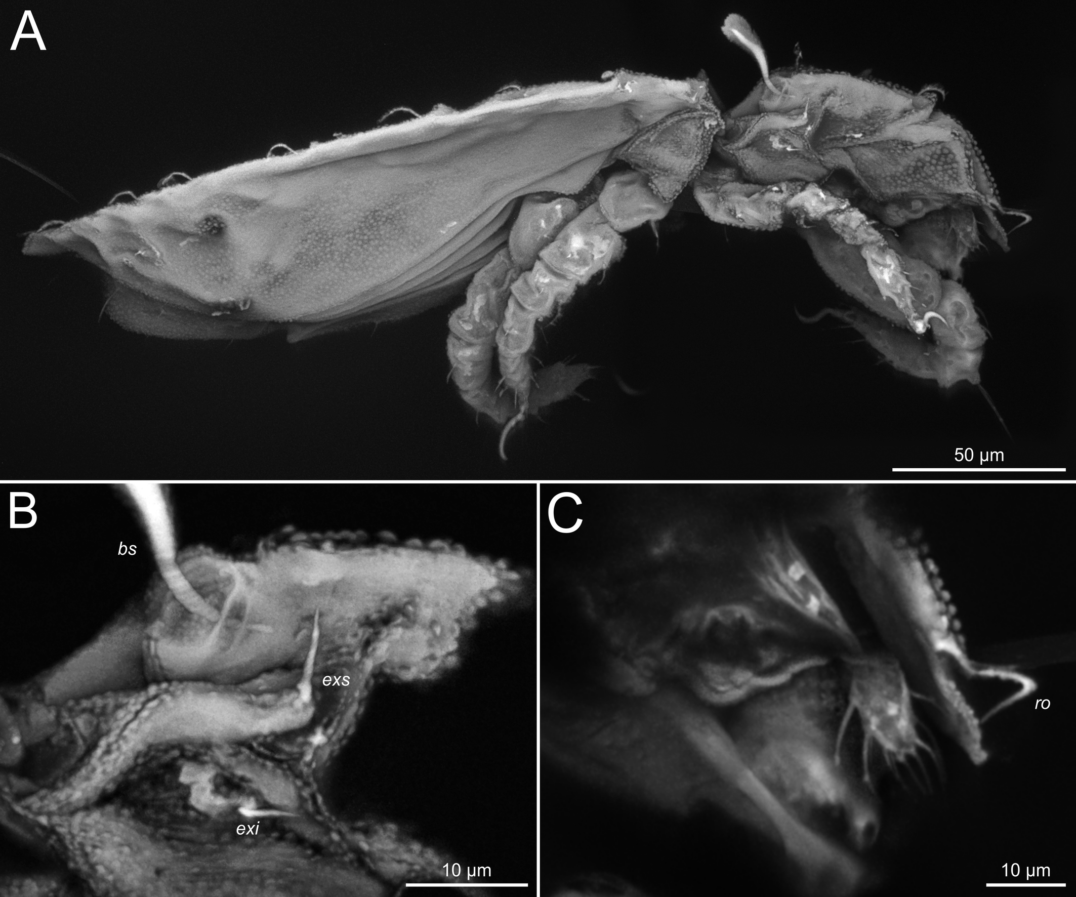
Integument — Where visible in transmitted light, parts of notogaster finely striated longitudinally (Fig. 1F); striation mostly hidden below cerotegument. Prodorsum and notogaster with large folds. Cerotegument microgranular: granules relatively large (about 1 µm), lentil-shaped. Smaller granules (about 0.5 µm and less) present around bothridia and at prodorsum-notogaster border (Fig. 2B, C, F). Cerotegumental layer extends beyond edges of cuticular shields (Figs. 2F, 3C).

Prodorsum — (Figs. 2A–E, 5B, 6A). Rostrum broadly rounded, with wide medial notch, without median apex, but with several sclerotized ridges. Rostral seta (ro, 25) curved inward, about equal in length to mutual distance, inserted on short apophyses, with rare barbs (Fig. 2D). Lamellar seta short (le, 13), not reaching insertion of rostral seta; with rare barbs, curved inward (Fig. 2E), mutual distance 27. Lamellar apophyses short (Fig. 2E). Bothridium funnel-shaped, directed posterolaterally (Figs. 2B, C, 5B). Bothridial seta long (bs, 45), club-shaped, finely barbed (Fig. 2B, C). Interlamellar seta short (in, 10) inserted medially close to bothridium, mutual distance 30. Exobothridial setae (exs, 10; exi, 8) inserted anterolateral to bothridium (Fig. 5A, B).

Notogaster — (Figs. 2A, 5A, 6A). Divided into four well defined shields by three transverse scissures. Notogaster with 15 pairs of short (not extending beyond the subsequent shield, except cp), smooth setae: shield Na with four pairs of setae (row c), c1 and c2 in central position, c3 and cp on either side near lateral margin, placed one posterior to the other (Fig. 2F); shield Nm1 with two pairs of setae (row d): one pair central and other pair lateral, inserted at mid-length of shield; shield Nm2 with two pairs of setae (row e), one pair central and other pair lateral, inserted close to anterior margin of shield; shield Py with seven pairs of setae (setal rows f, h and ps): setae f1, h1 and ps1 in central line and f2, h2 and ps2-3 close to dorsolateral margin (Fig. 2A, J). Seta ps3 positioned ventrally (Fig. 3A). Cupules not reliably and only unilaterally discerned (Fig. 2G), other cupules probably obscured by cerotegument.

Gnathosoma — (Figs. 3, 7E). Subcapitulum with three pairs of bifurcated adoral setae (or) (Figs. 3E, F). Ventral subcapitular setae (h, m, a) smooth. Pharyngeal cupola present. Palp five segmented; setal formula: 0-2-1-3-12(+ω); setae ul′ and ul′′ eupathidial (Fig. 3B). Chelicera with two setiform setae, cha and chb. For gnathosomal structures, see 3D reconstruction based on the CLSM image stack in the supplementary data.
Epimeral region — (Figs. 3A, 6B). Epimeres III and IV fused, epimeral setal formula: 3-2-2-3; all setae relatively short (16–27), thin and smooth.
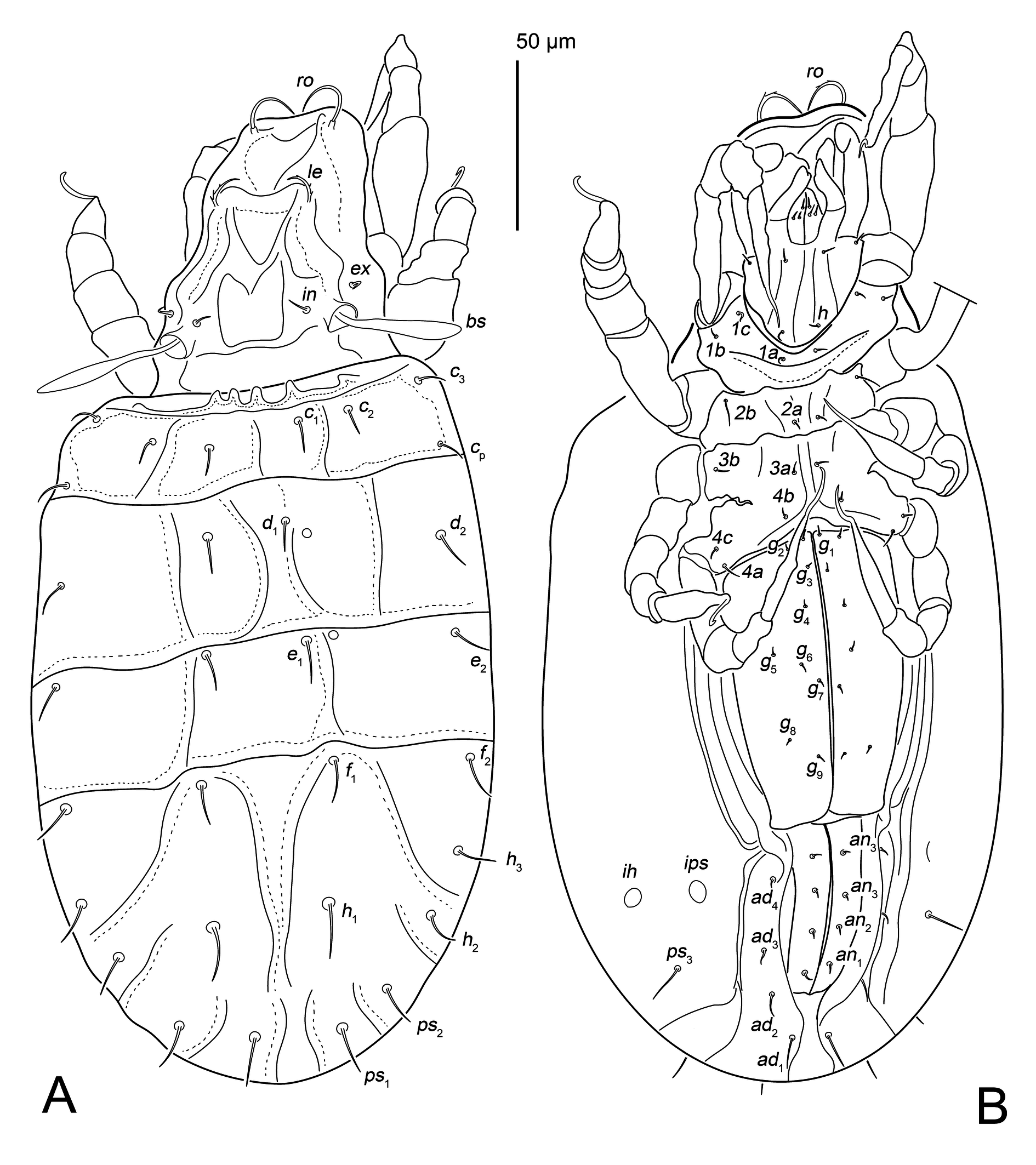
Ano-genital region — (Figs. 3D, 6B). Genital plates large (90 × 55), bearing nine pairs of smooth and thin setae; aggenital setae absent. Anal plates (55 × 23) with four pairs of smooth anal setae, adanal plates with four pairs of smooth adanal setae.

Legs — (Figs. 4, 7A–D). All legs monodactylous; claws slender, with proximoventral tooth at base (Fig. 4F). Leg chaetome (homologies of setae and solenidia indicated in Table 1): I 1-4-4-4(1)-17(1); II 1-4-4-4(1)-13(1); III 2-3-2-3(1)-12; IV 1-2-2-3-13. Solenidion ω1 of tarsus I slightly longer than famulus; solenidion φ I of tibia I much longer than short seta d; solenidion on tibia II–III shorter than respective seta d.
Download as Note: Roman letters refer to normal setae, Greek letters refer to solenidia (except ɛ = famulus); dφ –coupled seta and solenidion. Single prime (′) and double prime (′′) indicate setae on the anterior and posterior side of a segment, respectively. Parentheses indicate paired setae (′ and ′′).
Leg
Trochanter
Femur
Genu
Tibia
Tarsus
I
v′
bv′′, (l), d
v, (l), d
(v), l′, dφ
(pv), ft′, m′′, (a), s, ft′′, (tc), (it), (u), (p), ε, ω1
II
v′
bv′′, (l), d
v, (l), d
(v), l′, dφ
(pv), (a), s, ft, (tc), it′′, (u), (p), ω1
III
v′, l′
ev′, l′, d
l′, d
v′, l′, dφ
(pv), (a), s, ft, (tc), (u), (p)
IV
v′
ev′, d
l′, d
v′, l′, d
(pv), (a), s, (ft), (tc), (u), (p)
Remarks
State of preservation. The folds of prodorsum and notogaster, at least in some part, may be due to shrinking of the mite during fossilization. Interpretation of structures next to notogastral setae d1 and e1 as cupules (Fig. 2G) is questionable.
Status of Senilochthonius. Originally described as a separate genus (Mahunka 1992), Senilochthonius was later downgraded to subgeneric rank without comment (Subías 2004). Balogh and Balogh (2002) indicated the number of genital setae (9 vs. 7 or fewer pairs) as the distinguishing feature between Senilochthonius and Haplochthonius. These two characters reliably distinguish these two subgenera. Furthermore, Senilochthonius has a unique rostral shape (with a smooth medial incision vs. rounded in Haplochthonius) and ventral position of seta ps3 (vs. dorsal in Haplochthonius). All these characters are present in the fossil mite described here and allow its confident placement in subgenus Senilochthonius. Thus, we accept the subgeneric status of Senilochthonius.
Etymology
The new fossil species has been named in honor of Professor Roy Norton, an eminent acarologist who paid much of his research attention to the systematic relationships in one of his favorite groups, the enarthronote mites.

Discussion
Most of the records for Haplochthoniidae indicate that they can survive or may even prefer dry conditions. It is evident that they have evolved mechanisms to protect themselves from desiccation. According to Norton et al. (1983), one of the convergent traits in terrestrial acariform mites that is associated with adaptation to dry habitats is a regression of genital papillae – the structures that enable water absorption (Alberti 1979). Their absence in Haplochthoniidae, as well as in closely related Cosmochthoniidae, Pediculochelidae and in some other groups of Acari correlates with tolerance of dry environments. Mites of the family Pediculochelidae were repeatedly found in Myanmar amber (Kolesnikov et al. 2025; also D. Vorontsov, unpublished observation), which also suggests that dry conditions were present in the Cretaceous Myanmar amber forest, at least periodically. This is consistent with the study by Shi et al. (2022), which proposed the presence of xeromorphic plant communities and abundant, fire-prone vegetation there.
In the fossil mite studied here, we can see the presence of cerotegument, similar to that described by Bernini (1973) in Haplochthonius sanctaeluciae (see p. 481, Fig. I, plates 2, 3, 4). As many oribatid mites possess cerotegument, it is only natural that it is preserved during the fossilization in amber and can be properly studied with high resolution imaging. For example, on the legs of H. (Senilochthonius) nortoni sp. nov. (Fig. 3F), cerotegument looks similar to that described by Sidorchuk and Norton (2011) in their detailed re-description of fossil archaeorchestid mite Plategeocranus sulcatus (see their Figs. 38, 41, 44).
In many mites (Raspotnig & Matischek 2010) and in some other arthropods, such as whip spiders (Wolff et al. 2017), micro-granulated cerotegument provides hydrophobic properties of the body surface. At the same time, the inner layer of cerotegument may prevent desiccation (Raspotnig & Krisper 1998). Following this, it is natural to presume that H. (Senilochthonius) nortoni sp. nov. as well as extant Haplochthoniidae, and most other Protoplophoroidea, lived in dry microhabitats, which, at the same time, could be occasionally flooded. However, we cannot totally rule out the possibility that the fossil mite was entrapped in tree resin after falling from a tree hollow or from a bird's nest, given that extant H. simplex has been found inhabiting such places (Johnston, 1967).
The shape of the claw of H. (Senilochthonius) nortoni sp. nov. with the proximoventral tooth (Fig. 4F) suggests the use of two-point fixation on uneven or rounded surfaces during locomotion (Pfingstl 2023). As no extant species of Haplochthonius has been reported with a similar claw shape, it can be presumed that in the Cretaceous Kachin amber forest, these mites inhabited a different substrate that extant species are unlikely to inhabit today.
In conclusion, the mite described here, along with earlier discoveries of the fossils from the family Pediculochelidae (Kolesnikov et al. 2025), serves to reinforce the hypothesis that the Cretaceous Kachin amber forest was not a wet tropical rainforest but instead contained xeric or seasonally dry environments.
Acknowledgements
We are very grateful to Ilya Minaev (Ryazan, Russia) for donating the piece of amber from his private collection to our study, to Roy Norton (State University of New York, USA) for valuable suggestions during the preparation of the manuscript, to Evgeny Perkovsky (Natural History Museum of Denmark), Igor Shamshev (Zoological Institute, Russian Academy of Sciences) and Antonio Arillo (Complutense University of Madrid, Spain) for identification of syninclusions. We would like to thank the reviewers for corrections that improved the clarity of our paper. We acknowledge the use of Claude [claude.ai] for the language improvement and proofreading of the manuscript.
Funding
The research of VBK was conducted within the framework of State Assignment No. 124032500016-4. The work of DDV was conducted under the Institute of Developmental Biology, Government basic research program No. 0088-2024-0011. CLSM images were taken with the equipment of the Core Centrum of the Institute of Developmental Biology of the Russian Academy of Sciences.
References
- Alberti G. 1979. Fine structure and probable function of genital papillae and Claparede organs in Actinotrichida. In: Rodriguez J.G. (Ed.). Recent advances in acarology, Vol. II. New York: Academic Press. p. 501-507. https://doi.org/10.1016/B978-0-12-592202-9.50072-0
- Arillo A., Subías L.S., Huang D.Y. 2023. Oribatid mites in Burmese amber I. First record of the family Achipteriidae (Acariformes, Oribatida) in Cretaceous amber, with the description of a new species of Cerachipteria Grandjean, 1935. Palaeoentomology, 6: 443-446. https://doi.org/10.11646/palaeoentomology.6.5.1
- Balogh J., Balogh P. 2002. Identification keys to the oribatid mites of the extra-Holarctic regions, Vol. 1. Miskolc: Well-Press. pp. 453.
- Bernini F. 1973. Notulae oribatologicae. VII. Gli Oribatei (Acarida) dell′isolotto di Basiluzzo (Isole Eolie). Lavori della Società Italiana di Biogeografia, 3: 355-480. https://doi.org/10.21426/B63110608
- Coleman C.O. 2006. Substituting time-consuming pencil drawings in arthropod taxonomy using stacks of digital photographs. Zootaxa, 1360: 61-68. https://doi.org/10.11646/zootaxa.1360.1.4
- Elbadry E.A., Nasr A.K. 1979. A new oribatid mite from Egypt (Oribatei, Haplochthoniidae). Deutsche Entomologische Zeitschrift, 26(1-3): 85-87. https://doi.org/10.1002/mmnd.4800260109
- Fujikawa T. 1996. Oribatid mites from Picea glehnii forest at Mo-Ashoro, Hokkaido (12) A new species of the family Haplochthoniidae. Edaphologia, 56: 5-10.
- Grandjean F. 1947. Les Enarthronota (Acariens). Première série. Annales des Sciences Naturelles, Zoologie, 8: 213-248.
- Grandjean F. 1954. Les Enarthronota (Acariens). 4e série. Annales des Sciences Naturelles, Zoologie, 6(11): 311-335.
- Johnston D.E. 1967. Observations on Oribatei. Discovery of Haplochthonius simplex (Willmann) in North America (Acari-Acariformes). Proceedings of the Entomological Society of Washington, 69(4): 365.
- Kolesnikov V.B., Vorontsov D.D., Norton R.A., Klimov P.B. 2025. First fossil evidence of pediculochelid mites: two new species from Middle Cretaceous and Late Eocene amber revealing morphological stasis over at least 99 million years. Acarologia, 65(1): 67-90. https://doi.org/10.24349/uxz4-s4sq
- Mahunka S. 1992. New and interesting mites from the Geneva Museum LXIII. A survey of the oribatid fauna of Senegal (Acari: Oribatida). Revue Suisse de Zoologie, 99: 673-712. https://doi.org/10.5962/bhl.part.79846
- Mahunka S., Mejia-Recamier B.E. 1998. A new Haplochthonius Willmann, 1930 species from Mexico (Acari: Oribatida). Folia Entomologica Hungarica Rovartani Közlemények, 59: 267-270.
- Norton R.A. 2001. Systematic relationships of Nothrolohmanniidae, and the evolutionary plasticity of body form in Enarthronota (Acari: Oribatida). In: Acarology: proceedings of the 10th International Congress. Melbourne: CSIRO Publishing. p. 58-74.
- Norton R.A., Behan-Pelletier V.M. 2009. Suborder Oribatida. In: Krantz G.W., Walter D.E. (Eds). A manual of acarology. Lubbock: Texas Tech University Press. p. 430-564.
- Norton R.A., O'Connor B.M., Johnston D.E. 1983. Systematic relationships of the Pediculochelidae (Acari: Acariformes). Proceedings of the Entomological Society of Washington, 85: 493-512.
- Ordouni F., Akrami M.A., Ramroodi S. 2021. New species of primitive oribatid mite of the family Haplochthoniidae (Acari, Oribatida) from southeastern Iran. Systematic & Applied Acarology, 26(11): 2109-2117. https://doi.org/10.11158/saa.26.11.11
- Pepato A.R., Klimov P.B. 2015. Origin and higher-level diversification of acariform mites - evidence from nuclear ribosomal genes, extensive taxon sampling, and secondary structure alignment. BMC Evolutionary Biology, 15(1): 178. https://doi.org/10.1186/s12862-015-0458-2
- Pfingstl T. 2023. Sharp claws beneath our feet - the diversity of tarsal attachment devices of oribatid mites (Acari, Chelicerata, excluding Astigmata) - a review. International Journal of Acarology, 49(3-4): 165-195. https://doi.org/10.1080/01647954.2023.2223214
- Rasnitsyn A.P., Bashkuev A.S., Kopylov D.S., Lukashevich E.D., Ponomarenko A.G., Popov Yu.A., Rasnitsyn D.A., Ryzhkova O.V., Sidorchuk E.A., Sukatsheva I.D., Vorontsov D.D. 2016. Sequence and scale of changes in the terrestrial biota during the Cretaceous (based on materials from fossil resins). Cretaceous Research, 61: 234-255. https://doi.org/10.1016/j.cretres.2015.12.025
- Raspotnig G., Krisper G. 1998. Fatty acids as cuticular surface components in oribatid mites (Acari: Oribatida). In: Ebermann E. (Ed.). Arthropod biology: contributions to morphology, ecology and systematics. Biosystematics and Ecology Series 14. p. 215-243.
- Raspotnig G., Matischek T. 2010. Anti-wetting strategies of soil-dwelling Oribatida (Acari). Acta Societatis Zoologicae Bohemicae, 74: 91-96.
- Sanyal A.K., Basak S., Barman R.P. 2002. Three new species of oribatid mites (Acarina, Oribatida: Haplochthoniidae) from the Antarctic continent. Acarina, 10(1): 57-63.
- Schindelin J., Arganda-Carreras I., Frise E., Kaynig V., Longair M., Pietzsch T., Preibisch S., Rueden C., Saalfeld S., Schmid B., Tinevez J.-Y., White D.J., Hartenstein V., Eliceiri K., Tomancak P., Cardona A. 2012. Fiji: an open-source platform for biological-image analysis. Nature Methods, 9(7): 676-682. https://doi.org/10.1038/nmeth.2019
- Sellnick M. 1931. Milben im Bernstein. Bernstein-Forschung, 2: 148-180.
- Shi G., Grimaldi D.A., Harlow G.E., Wang J., Wang J., Yang M., Lei W., Li Q., Li X. 2012. Age constraint on Burmese amber based on U-Pb dating of zircons. Cretaceous Research, 37: 155-163. https://doi.org/10.1016/j.cretres.2012.03.014
- Shi C., Wang S., Cai H.H., Zhang H.R., Long X.X., Tihelka E., Song W.C., Feng Q., Jiang R.X., Cai C.Y., Lombard N. 2022. Fire-prone Rhamnaceae with South African affinities in Cretaceous Myanmar amber. Nature Plants, 8(2): 125-135. https://doi.org/10.1038/s41477-021-01091-w
- Sidorchuk E.A., Norton R.A. 2011. The fossil mite family Archaeorchestidae (Acari, Oribatida) II: redescription of Plategeocranus sulcatus and family-group relationships. Zootaxa, 3051(1): 14-40. https://doi.org/10.11646/zootaxa.3051.1.2
- Sidorchuk E.A., Vorontsov D.D. 2018. Preparation of small-sized 3D amber samples: state of the technique. Palaeoentomology, 1(1): 80-90. https://doi.org/10.11646/palaeoentomology.1.1.10
- Smith R.D., Ross A.J. 2016. Amberground pholadid bivalve borings and inclusions in Burmese amber: implications for proximity of resin-producing forests to brackish waters, and the age of the amber. Earth and Environmental Science Transactions of the Royal Society of Edinburgh, 107(2-3): 239-247. https://doi.org/10.1017/S1755691017000287
- Subías L.S. 2004. Listado sistemático, sinonímico y biogeográfico de los ácaros oribátidos (Acariformes: Oribatida) del mundo (excepto fósiles). Graellsia, 60: 3-305. https://doi.org/10.3989/graellsia.2004.v60.iExtra.218
- Subías L.S. 2022. Listado sistemático, sinonímico y biogeográfico de los ácaros oribátidos (Acariformes, Oribatida) del mundo (excepto fósiles) [Internet]. Monografías Electrónicas, 12: 1-538. Available from: http://sea-entomologia.org/MeSEA_12_Listado_mundial_Acaros_Oribatidos_L_Subias.pdf. Accessed 27 March 2026.
- Subías L.S., Arillo A. 2002. Oribatid mite fossils from the Upper Devonian of South Mountain, New York and the Lower Carboniferous of County Antrim, Northern Ireland (Acariformes, Oribatida). Estudios del Museo de Ciencias Naturales de Alava, 17: 93-106.
- Suzuki K. 1977. Two species of Enarthronota: Haplochthonius simplex and Cosmochthonius reticulatus. Bull. Biogeogr. Soc. Jap., 32(2): 5-16.
- Weigmann G. 2006. Hornmilben (Oribatida). Die Tierwelt Deutschlands, Teil 76. Keltern: Goecke & Evers. pp. 520.
- Willmann C. 1930. Neue und bemerkenswerte Oribatiden aus der Sammlung Oudemans. Abhandlungen des Naturwissenschaftlichen Vereins zu Bremen, 28(1): 1-12.
- Wolff J.O., Seiter M., Gorb S.N. 2017. The water-repellent cerotegument of whip-spiders (Arachnida: Amblypygi). Arthropod Structure & Development, 46(1): 116-129. https://doi.org/10.1016/j.asd.2016.10.010
to post any comment or correction.


2026-04-02
Date accepted:
2026-07-06
Date published:
2026-07-09
Edited by:
Baumann, Julia

This work is licensed under a Creative Commons Attribution 4.0 International License
2026 Vorontsov, Dmitry D. and Kolesnikov, Vasiliy B.
 Download article Download low definition
Download the citation
RIS with abstract
(Zotero, Endnote, Reference Manager, ProCite, RefWorks, Mendeley)
RIS without abstract
BIB
(Zotero, BibTeX)
TXT
(PubMed, Txt)