


New French tiny spider mites (Prostigmata, Tetranychidae) on a tiny broom
Auger, Philippe  1
; Arabuli, Tea
2
and Migeon, Alain
3
1
; Arabuli, Tea
2
and Migeon, Alain
3
1✉ CBGP, Univ Montpellier, CIRAD, INRAE, IRD, Montpellier SupAgro, Montpellier, France.
2Institute of Zoology, Ilia State University, Kakutsa Cholokashvili Ave 3/5, Tbilisi 0162, Georgia & Institute of Entomology, Agricultural University of Georgia, Kakha Bendukidze Campus, 240 David Aghmashenebeli Alley, 0131, Tbilisi, Georgia.
3CBGP, Univ Montpellier, CIRAD, INRAE, IRD, Montpellier SupAgro, Montpellier, France.
2022 - Volume: 62 Issue: 3 pages: 672-693
https://doi.org/10.24349/e34b-1nnyZooBank LSID: 918203DC-09E4-45AC-8F77-26F6BC0AA8D6
Original research
Keywords
Abstract
Introduction
According to the number of recorded species, Europe has the greatest specific diversity for the genus Bryobia with about 45% of the recorded species (Migeon and Dorkeld, 2006-2021) and of 70% of the species from the Palearctic region. This trend is confirmed by the location of the newly described species belonging to this genus in the last ten years. During this period, of the seven newly described species worldwide, six were from the palearctic region: two from Syria (Barbar and Auger, 2020; Barbar et al., 2022) and four from Europe, in France (Auger and Migeon, 2014; Auger et al., 2015). As a result, the genus Bryobia is very diversified in Europe with 54 recorded species (see Migeon and Dorkeld, 2006-2021).
The work by Eyndhoven and Vacante (1985) on the Bryobia species belonging to the berlesei-group and some more recent works (Auger and Migeon, 2014; Auger et al., 2015, Barbar et al. 2022) have shown that the plants of the family Fabaceae, from the tribe Genisteae, belonging to the genera Cytisus (including Calicotome), Genista (including Teline and Retama), and Ulex, are favourable to many Bryobia species. In France, these plant genera are quite well represented with 53 species recorded (Coulot and Rabaute, 2016) and several of them remain unsampled for spider mite detection.
Therefore, when the last author remembered that during his studies, the French botanist Dr Yves Baron showed him the tiny broom Genista pulchella Vis. [French name: ''genêt joli'' meaning ''pretty broom'', (Fig. 1)] in a location quite close to our laboratory, we wanted to know if, despite its very reduced size, some Bryobia mites could inhabits this plant.

The sampling events of G. pulchella we undertook in several locations in southern France disclosed two unknown spider mite species: one belonging to the genus Bryobia but also one belonging to the genus Tenuipalpoides. The aim of the present work is to provide the descriptions of these two new species. In addition, the observation of type specimens of Tenuipalpoides zizyphus Reck and Bagdasarian, 1948 for comparison with the new Tenuipalpoides species allowed us to provide updated body measurements (those given in the original description were smaller than in reality) and some additional morphological data that were missing or inaccurate in the original description.
Material and methods
Mites were extracted from broom using the washing method (Boller, 1984), by filtering the solution on three stacked sieves (smallest mesh size 400), and picking out mites using a camel hair brush. They were preserved in 70% ethyl alcohol. Following clearing in lactic acid (50%) for 24 hours, mites were mounted in Hoyer's medium. The specimens were examined using a Leica® DM LB2 phase contrast microscope. Drawings were made with a Wacom Intuos Pro L pen tablet using stacks of photographs acquired from the AmScope® MU1803 camera and imported to Adobe Illustrator® CS5. Line drawings were vectorised, edited and placed into figures using Adobe Illustrator® CS5. Measurements were taken with live images using the software Amscope® suite (v. 3.7.7934) coupled with the above mentioned camera.
The setal nomenclature used in the description follows Lindquist (1985). Leg setal counts are given according to the sequence coxa-trochanter-femur-genu-tibia-tarsus. Numbers of setae refer to tactile setae, solenidia are given in parentheses. The most frequent number of setae in a leg segment is provided first and alternative counts are given in brackets. All measurements are presented in micrometres (µm) and correspond to the holotype followed (in parentheses) by minimum and maximum values from paratypes. Body length measurement represents the distance between the tip of gnathosoma (when including gnathosoma) or the tip of inner prodorsal lobes (when excluding gnathosoma) to the caudal end of idiosoma and width represents the widest transversal part of the hysterosoma. Distance between sc2 setae members and between v2 and h1 setae are also given (Saito et al., 1999). Setae were measured from the centre of their setal bases to their tips. The distance between two setae was measured as the distance from the centre of one setal base to the other. In bryobiine mites, the inner lobe height was measured as the orthogonal distance from the bottom of outer incisions to the tip of the inner lobes (excluding setae v1). Prodorsal lobes basal width was measured as the distance between the external margin of the bases of the external lobes. The following abbreviations are used for institutions: CBGP = Centre de Biologie pour la Gestion des Populations; INRAE = Institut National de Recherche pour l′Agriculture, l′Alimentation et l′Environnement. All the mite specimens are deposited in the Centre de Biologie pour la Gestion des Population. 2018. CBGP - Continental Arthropod Collection. https://doi.org/10.15454/D6XAKL ![]()
Results and discussion
Family Tetranychidae Donnadieu, 1875
Subfamily Bryobiinae Berlese, 1913
Tribe Bryobiini Reck, 1952
Genus Bryobia Koch, 1836
Bryobia Koch, 1836 :8-9 Type-species: Bryobia praetiosa Koch.
Bryobia (Lyobia) baroni sp. n.
ZOOBANK: 33013FE0-200F-4C9A-8181-0267155785A2 ![]()
(Figures 2-5)
Type material
Holotype (female) — 11 female, 8 deutonymph, 1 protonymph paratypes on 21 microscopic preparations from Genista pulchella Vis. (Fabaceae), Top of Mont Tauch (42.9096°N 2.6794°E, 840 m a.s.l.), Tuchan (Aude), France, 18-IX-2014, leg. A. Migeon and P. Auger; 10 female, 2 protonymph, 7 larva paratypes on 17 microscopic preparations from G. pulchella, Le Jouquet, forêt domanaliale de Partlages (43.7746°N 3.4394°E, 770 m a.s.l.), Saint-Pierre-de-la-Fage (Hérault), France, 26-VII-2012, leg. A. Migeon and P. Auger. Collection Auger-Migeon N° 1864 for holotype, 1865–1901 for paratypes.
Additional material — 5 females, one nymph on 6 preparations from Genista pulchella Vis. (Fabaceae), Top of Mont Tauch (42.9097°N 2.6794°E, 840 m a.s.l.), Tuchan (Aude), France, 25-VI-2012, leg. A. Migeon and P. Auger; 1 nymph, 1 larva on 2 microscopic preparations from G. pulchella, La Crémade on D94 (44.2563°N 3.0669°E, 889 m a.s.l.), Sévérac-le-Château (Aveyron), France, 19-VII-2012, leg. A. Migeon and P. Auger; 11 females, 4 nymphs on 15 microscopic preparations from G. pulchella, Piste des Indochinois (44.0128°N 5.2981°E, 779 m a.s.l.), Méthamis (Vaucluse), France, 25-IX-2014, leg. A. Migeon and P. Auger; 12 females, one nymph on 11 preparations from Genista pulchella Vis. (Fabaceae), Peyrafic – Route des Cèdres, Parc Naturel du Luberon (43.8049°N 5.2871°E, 635 m a.s.l.), Bonnieux (Vaucluse), France, 25-IX-2014, leg. A. Migeon and P. Auger.
Diagnosis
This species belongs to the subgenus Lyobia having prodorsal lobes well developed, no duplex setae on tarsus IV (Livshits and Mitrofanov, 1971) and to the berlesei-group (all living on Genisteae) due to a femoral interior dorsal row of leg I bearing four long setae (Eyndhoven and Vacante, 1985). Females with body small, legs shorter than body length; anterior dorsal prodorsal projections over gnathosoma well developed, outer prodorsal lobes conical, inner lobes triangularly shaped well separated by ''u'' or v-shaped incision, outer incisions wider; dorsal body setae spatulate, serrate, short, subequal in length, inserted on small bulge-like structures becoming obvious tubercles caudally; among dorsocentral setae, c1 the largest, d1 and e1 the shortest; palptarsus far longer than bidentate tibial claw; spermatheca sacculus oval; empodia I with a pair of tenent hairs, others with two rows of tenent hairs; tarsus IV with 14 setae and one solenidion, femur IV with 5 setae.
Description


Female — (Figs. 2-5). Holotype 530 long (excluding gnathosoma), 630 (including gnathosoma), distance between setae v2-h1 510, width 380, distance between setae sc2 members 285; 13 paratypes measured, 506-596 long (excluding gnathosoma), 575-675 (including gnathosoma), distance between setae v2-h1 470-565, width 345-390, distance between setae sc2 members 260-297.
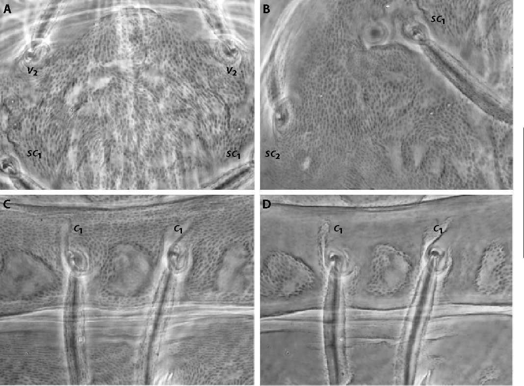
Dorsum – Prodorsum with four pairs of setae with well-developed anterior prodorsal lobes (Figs. 2, 3A-B). Prodorsal lobes with basal width about 105 (90-105), outer prodorsal lobes conical, inner lobes triangularly shaped, 32 (31-44) high, usually well separated by bottom U-shaped incision, sometimes varying to V-shaped, 14 (11-20) in depth (Fig. 3A-B). Incisions between median and outer lobes wider, bottom rounded. Propodosomal setae v2 of outer lobes reaching or slightly extending beyond the base of setae v1 of inner lobes. Distance between v1 and v2 setae insertions 25 (15-30) and 88 (73-89), respectively; setae v1 and v2 spatulate elongate, v2 setae wider: 7 (6-8) and 10 (9-13) in width, respectively. Dorsal body setae spatulate, rounded distally, rough, serrate, subequal in length, inserted on small bulge-like structures becoming obvious tubercles caudally, f1 and f2 setae in marginal position not contiguous (Fig. 2A-C). Dorsocentral setae (c1, d1 and e1) shorter than distances between consecutive setae, c1 the largest, d1 and e1 the shortest (Fig. 2B-C): v1 20 (17-29); v2 30 (26-39); sc1 28 (22-31); sc2 24 (20-27); c1 26 (25-29); c2 26 (22-27); c3 22 (20-25); d1 22 (19-24); d2 21 (20-24); d3 24 (21-24); e1 23 (20-24); e2 24 (21-25); e3 22 (21-28); f1 22 (21-29); f2 26 (22-30); h1 25 (21-32). Distances between setae: c1-c1 61 (63-76), d1-d1 57 (45-61), e1-e1 35 (26-37), c3-c3 313 (286-326), c1-d1 83 (70-94), d1-e1 70 (57-84), v1-v1 25 (15-30), v2-v2 88 (73-89), sc2-sc2 287 (260-300). Dorsal integument on propodosoma with irregular discontinuous folds forming more or less an oval pattern with longitudinal folds medially becoming oblique laterally (Figs. 2, 3C); prodorsal medial folds integument with irregular stout dashed striation (Fig. 3C). Hysterosomal folds mainly transverse with three pairs of more or less oval-shaped areas with irregular folds present between setae c1- c2, d1- d3, and e1- e3 and one caudally (Fig. 2). Hysterosomal medial folds integument near setae c1 with irregularly dashed fine striation becoming finely striated near setae d1 and e1 (Fig. 3D).
Gnathosoma – Stylophore notched longer than wide 120 (115-125) and 75 (70-90), respectively. Palptarsus well-developed 40 (33-39) (including eupathidia), 31 (26-32) (without eupathidia), far longer than bidentate tibial claw, tip of tibial claw do not reach insertion of proximal palptarsal tactile seta a, palptarsus with three tactile setae, three eupathidia and one solenidion (Fig. 3E): eupathidia ul′ζ, ul″ζ 10 (9-10) slightly shorter than solenidion 11 (10-12), suζ shorter 6 (5-7). Peritreme anastomosed distally in a relatively long and slender enlargement, 38 (30-40) long, 8 (7-8.5) wide (Fig. 3F).
Venter – Striation transverse between 1st pair of setae (1a) and second pair (3a), almost smooth between setae 3a and 4a, transverse posteriorly to setae 4a becoming longitudinal from aggenital (ag); area immediately anterior to genital opening with irregular longitudinal striation (Fig. 3H). Two pairs of ventrocaudal (h2-3) setae and three pairs of pseudanal setae (ps1-3) present. Spermatheca oval in shape 11 (10-11) long, 7 (6.5-7) wide (Fig. 3G).
Legs – Legs shorter than body length. Leg I 420 (385-425) long (measured from femur to tarsus) about ¾ body length, longer than leg II 240 (210-240), leg III 240 (215-245) and leg IV 280 (240-280). Length of segments of leg I as follows: trochanter 50 (40-50), femur 150 (130-155), genu 65 (65-75), tibia 105 (95-105), tarsus 100 (90-105). Tibia I and tarsus I about 2/3 length of femur I. Leg setal counts as follows (Fig. 4A-D):
I 2 − 1 – 12 [11-14] – 7 [5-8] – 13 + (1) – 19 + (5) + 2 duplexes;
II 1 − 1 – 9 [7-8] – 5 – 9 [7-8] – 15 + (2) + 1 duplex;
III 1 − 1 – 6 [7] – 6 [5-7] − 9 – 13 + 1 duplex;
IV 1 − 1 – 5 [4] – 6 − 9 – 14 + (1).
Femur I usually bearing 4 (sometimes 3, rarely 2, on one of the two femora I) large serrate elongate setae (subproximal seta bl and 3 setae l) in the internal femoral upper row, the longest about 40 (37-44). Empodium I with one pair of tenent hairs, other empodial pads each provided with two rows (Fig. 4E-F). True claws uncinate each with one pair of tenent hairs on leg I, other claws usually with two/three pairs but sometimes four pairs present (Fig. 4E-F). Coxisternal seta 1b slender smooth 49 (40-50), coxisternal seta 1c shorter 18 (17-30), smooth to finely serrate (Fig. 4G). Solenidion on tibia I 15 (12-17). Tarsus III associated setae approximate with solenidion forming duplex, tactile member (17-21) slightly serrate dorsally, proximal, slightly longer than solenidion (13-18) (Fig. 4C); tarsus IV with solenidion 10 (8-10) well-separated from tactile 20 (18-20), proximal, about half the length of tactile (Fig. 4D). Tectal setae (tc′-tc″) smooth, elongated, slender on tarsus I, slightly serrate and furcate distally on other tarsi.
Deutonymph — (4 paratypes measured) (Fig. 5A). Body 430-470 long (excluding gnathosoma), 520-540 long (including gnathosoma), distance between setae v2 – h1 430-450, width 290-315, distance between sc2 members 220-240.
Dorsum – Prodorsal lobes developed, outer small more or less triangularly shaped, inner lobes more developped, triangular, 20-24 high, well separated by v-shaped incision 7-10 in depth. Prodorsal setae v2 of outer lobes reach about 2/3 of setae v1, v2 about 1.5 times the length of v1. Dorsal setae inserted on small tubercles (except setae v1 and v2), more obvious on hysterosoma and stronger caudally. Prodorsal setae serrate, spatulate, hysterosomal setae serrate, setae c1 to e2 spatulate, setae e2 narrower and longer, setae e3 subspatulate, other caudal setae f1, f2 and h1, serrate, subspatulate to elongate wide, more or less rounded distally (Fig. 5A).
Dorsocentral setae the shortest, f1, f2 and h1 the longest: v1 15-18; v2 26-29; sc1 25-29; sc2 18-23; c1 22-24; c2 18-22; c3 19-23; d1 19-21; d2 20-23; d3 22-27; e1 19-20; e2 20-28; e3 25-34; f1 29-44; f2 32-38; h1 22-37. Distances between setae: c1-c1 58-60, d1-d1 40-45, e1-e1 27-32, c1-d1 75-84, d1-e1 60-72, v1-v1 16-17, v2-v2 57-65.
Gnathosoma – Stylophore slightly emarginate, longer than wide, 85-95 and 60, respectively. Peritreme anastomosed distally in a relatively long and slender enlargement, 15-20 long, 6-7 wide. Palptarsus 29-30 long (including eupathidia) far longer than bidentate tibial claw, tip of tibial claw do not reach insertion of proximal palptarsal tactile seta a.
Legs – Length inferior to body length, leg I 230-255 long (from femur to tarsus), about 1.5 longer than leg II 160-180, leg III 160-170, leg IV 170-185. Leg setal counts as follows:
I 2 − 1 – 8 − 4 − 9 + (1) – 14 +(1) + 2 duplexes;
II 1 − 1 – 6 − 4 − 5 – 11[10] + 1 duplex;
III 1 − 1 – 2– 3 − 5 – 9 + 1 duplex [10+(1)];
IV 1 − 0 – 2 – 3 − 5 – 10.
Duplex on tarsus III sometimes dissociate on one side of the mite. Empodial pads each provided with two rows of tenent hairs. True claws uncinate with one pair of tenent hairs, rarely two pairs on claws II, sometimes two pairs on claws III-IV. On femur I, two serrate macrosetae present, the longest 28-32.
Protonymph — (3 paratypes measured) (Fig. 5B). Body 295-325 long (excluding gnathosoma), 345-390 long (including gnathosoma), distance between setae v2 – h1 280-310, width 200-210, distance between sc2 members 155-175.
Dorsum – Prodorsal lobes poorly developed, outer very small, inner small triangular, separated by wide weak incision 4 in depth. Prodorsal setae v2 surpass well tip of setae v1, v2 about 2.5 times the length of v1. Dorsal setae serrate inserted on tiny tubercles caudally. Setae c1, c2, d1, d2 and e1 spatulate, d3 and e2 subspatulate, other caudal setae elongate, narrower, acute distally (Fig. 5B). On hysterosoma, setae c2 and c3 the shortest, setae e2 to h1 the longest: v1 10-11; v2 24-26; sc1 22-25; sc2 18-19; c1 19-20; c2 16-19; c3 16-16; d1 16-17; d2 20-22; d3 24-26; e1 19-20; e2 26-31; e3 31-34; f1 28-34; f2 31-33; h1 29-32. Distances between setae: c1-c1 50-60, d1-d1 35-39, e1-e1 22-25, c1-d1 48-55, d1-e1 36-48, v1-v1 11, v2-v2 45-47.
Gnathosoma – Stylophore very slightly emarginate, longer than wide, 75-85 and 60-70, respectively. Peritreme anastomosed distally in slender enlargement, 17-19 long, 4 wide. Palptarsus 19-25 long (including eupathidia) far longer than bidentate tibial claw, tip of tibial claw do not reach insertion of proximal palptarsal tactile seta a.
Legs – Length inferior to body length, leg I 165-185 long (from femur to tarsus), leg II 120-130, leg III 110-130, leg IV 115-130. Leg setal counts as follows:
I 2 − 0 – 3 − 4 − 5 + (1) – 10 + 2 duplexes;
II 1 − 0 – 3 − 4 − 5 – 9 + 1 duplex;
III 1 − 0 – 2– 2 − 5 – 8;
IV 0 − 0 – 2 – 2 − 5 – 6.
Empodial pads each provided with two rows of tenent hairs. True claws uncinate with one pair of tenent hairs. On femur I, one serrate macrosetae present 23-24 long.
Larva — (4 paratypes measured) (Fig. 5C). Body 305-330 long (including gnathosoma), distance between setae v2 – h1 230-250, width 185-190, distance between sc2 members 135-140.
Dorsum – Prodorsal lobes absent. Prodorsal setae v2 surpass well tip of setae v1, v2 about 3 times the length of v1. Dorsal setae elongate serrate acute distally, inserted on tiny bulges posteriorly. Setae c1, c2, d1, d2 and e1 slightly wider (Fig. 5C).
On hysterosoma, setae c2-3 and d2 the shortest, setae f1-2 and h1 the longest: v1 6-8; v2 21-23; sc1 19-20; sc2 15-19; c1 20-24; c2 16-19; c3 15-18; d1 19-23; d2 16-19; d3 21-24; e1 24-25; e2 22-27; e3 27-29; f1 27-33; f2 28-35; h1 27-31. Distances between setae: c1-c1 57-64, d1-d1 28-31, e1-e1 16-19, c1-d1 49-55, d1-e1 39-40, v1-v1 10-11, v2-v2 43-45.
Gnathosoma – Stylophore very slightly emarginate, longer than wide, 60-70 and 50-55, respectively. Distal end of peritreme not seen. Palptasus 22-24 long (including eupathidia) far longer than bidentate tibial claw, tip of tibial claw reach insertion of proximal palptarsal tactile seta a.
Legs – Length inferior to body length, leg I 130-140 long (from femur to tarsus), leg II 105-115, leg III 105-110. Leg setal counts as follows:
I 1 − 0 – 3 − 4 − 5 + (1) – 7 + 1 duplex;
II 0 − 0 – 3 − 4 − 5 – 7 + 1 duplex;
III 0 − 0 – 2– 2 − 5 – 6.
Empodial pads shorts with 2/3 pairs of tenent hairs on leg I, pads longer on legs II-III with 2 rows of tenent hairs. True claws uncinate with one pair of tenent hairs. On femur I, one serrate macrosetae present 15-18 long.
Male — Unknown
Remarks
Among the species belonging to the berlesei-group , B. (L.) baroni n. sp. is closely related to B. (L.) provincialis Van Eyndhoven and Vacante, 1985 and more to B. (L.) dikmenensis Van Eyndhoven and Vacante, 1985 by having short dorsal setae, 4 long and 1 normal setae on femur I, interior lobes more developed than outer ones, empodium I with a pair of tenent hairs. It can be easily separated from B. (L.) provincialis by the following characters: 1) it is smaller in size (in the new species length 506-596 and width 345-390 versus 560-740 and 410-600 in B. (L.) provincialis); 2) different inner lobes that are triangular and well separated by a ''v'' or u-shaped incision in the new species vs. mammelliform and largely fused in B. (L.) provincialis; 3) two small differences in setal count on femur and tarsus IV, 5 and 14+(1) setae, respectively in B. (L.) baroni n. sp. vs. 6 and 13+(1) in B. (L.) provincialis; 4) the first pair of dorsocentral setae (c1) is obviously larger than the second and the third pairs (d1, e1) in the new species vs. setae c1, d1 and e1 of similar size in B. (L.) provincialis; 5) a longer palptarsus with the palpal claw not reaching the proximal palpal tactile seta a in the new species vs. a palpal claw reaching the proximal palpal tactile seta in B. (L.) provincialis and 6) a shorter leg I in the new species, 385-425 long vs. 446-650 in B. (L.) provincialis.
Despite a body lenght similar, females of B. (L.) baroni n. sp. differ from B. (L.) dikmenensis by: 1) a body less narrow in the new species about 355-390 vs. 250-315 in B. (L.) dikmenensis; 2) inner lobes triangularly shaped, not inflated, well separated by bottom ''v'' or u-shaped incision in B. (L.) baroni n. sp. vs. inner lobes mammelliform and largely fused in B. (L.) dikmenensis; 3) a larger distal peritremal anastomosis in B. (L.) baroni n. sp. than in B. (L.) dikmenensis, 32-40 and 29-30, respectively; 4) a larger stylophore in B. (L.) baroni n. sp. than in B. (L.) dikmenensis, 115-125 x 70-85 and 100 x 55, respectively; 5) longer legs II, III and IV in B. (L.) baroni n. sp. than in B. (L.) dikmenensis, 225-245, 235-245, 250-280 and 200, 200, 220, respectively and 6) longer large setae on femur I in B. (L.) baroni n. sp. than in B. (L.) dikmenensis, 38-43 and 36, respectively.
Etymology
The species designation ''baroni'' refers to the French Botanist Dr Yves Baron, recently deceased, who showed to the last author, during his studies, some Genista pulchella at the top of the Mont Tauch (Aude, Southern France) (Fig. 1).
Subfamily Tetranychinae Berlese, 1913
Tribe Tenuipalpoidini Pritchard and Baker, 1955
Genus Tenuipalpoides Reck and Bagdasarian, 1948
Tenuipalpoides Reck and Bagdasarian, 1948: 183-186. Type-species: Tenuipalpoides zizyphus Reck and Bagdasarian.
Tenuipalpoides genistearum sp. nov.
ZOOBANK: 21D1E64D-D205-40C1-8A93-CF7E5363EFD2 ![]()
(Figures 6-12)


Type-material
Holotype (female) — 10 female, 2 male, 9 nymph, 1 larva paratypes on 20 microscopic preparations from Genista pulchella Vis. (Fabaceae), Camp Gourens, Znieff 9010011840 (43.8824°N 3.5566°E, 559 m a.s.l.), Rogues (Gard), France, 26-VII-2012, leg. A. Migeon and P. Auger; 1 male paratype on 1 microscopic preparations from G. pulchella, Le Jouquet, forêt domaniale de Notre Dame de Parlatges (43.7746°N 3.4394°E, 770 m a.s.l.), Saint-Pierre-de-la-Fage (Hérault), France, 26-VII-2012, leg. A. Migeon and P. Auger; 8 female, 5 male, 3 nymph paratypes on 16 microscopic preparations from G. pulchella, Piste des Indochinois (44.0128°N 5.2981°E, 779 m a.s.l.), Méthamis (Vaucluse), France, 25-IX-2014, leg. A. Migeon and P. Auger. Collection Auger-Migeon N° 1902 for holotype, 1903–1938 for paratypes.
Additional material — 4 females, 10 nymphs, 1 larva on 14 microscopic preparations from G. pulchella, Col d'Engayresque on D294 (44.2539°N 3.0617°E, 873 m a.s.l.), Verrières (Aveyron), France, 19-VII-2012, leg. A. Migeon and P. Auger; 12 females, 2 nymphs, 1 larva on 15 microscopic preparations from G. pulchella, La Crémade on D94 (44.2564°N 3.0669°E, 889 m a.s.l.), Sévérac-le-Château (Aveyron), France, 19-VII-2012, leg. A. Migeon and P. Auger; 8 females, 6 nymphs, 1 larva on 15 microscopic preparations from G. pulchella, Le Jouquet, forêt domaniale de Notre-Dame-de-Parlatges (43.7746°N 3.4394°E, 770 m a.s.l.), Saint-Pierre-de-la Fage (Hérault), France, 26-VII-2012, leg. A. Migeon and P. Auger; 10 females, 3 males, 6 nymphs deutonymph on 19 microscopic preparations from Genista pulchella Vis. (Fabaceae), Top of Mont Tauch (42.9097°N 2.6794°E, 840 m a.s.l.), Tuchan (Aude), France, 18-IX-2014, leg. A. Migeon and P. Auger.
Diagnosis
Females with dorsal setae oblong inserted on strong tubercles; dorsal integument finely granulated, covered by tiny dense pimple-like bulges; propodosoma with small irregular shallow dimples forming an irregular not meshlike and blurred alveoli pattern medially; deeper irregularly shaped depressions present on dorsal hysterosomal integument; peritremal distal end a simple hook rarely branched, leg I tarsal setal count very reduced. Male aedeagus bent dorsad near at a right angle, bent part without knob, almost straight, progressively tapering.
Description
Female — (Figs. 6-9). Holotype 355 long (excluding gnathosoma) 415 (including gnathosoma), distance between setae v2-h1 340, width 260, distance between setae sc2 members 205; 9 paratypes measured, 325-360 long (excluding gnathosoma) 400-430 (including gnathosoma), distance between setae v2-h1 310-340, width 230-280, distance between setae sc2 members 190-225.
Dorsum – All dorsal body setae set on strong tubercles, larger caudally, v2 tubercles the smallest. Dorsal setae oblong, variable in size and shape according to their dorsal location, serrate with rachis-like stem on the lower surface (Fig. 6A-C). First and second pair of dorsocentral hysterosomal setae (c1, d1) reach insertions of consecutive setae. Dorsal setal lengths: v2 56 (54-61); sc1 55 (55-65); sc2 48 (41-53); c1 64 (58-64); c2 62 (60-69); c3 55 (43-54); d1 64 (62-70); d2 63 (58-67); e1 66 (63-69); e2 62 (58-67); f1 58 (54-65); f2 59 (54-66); h1 58 (54-61). Distances between seate: c1-c1 39 (38-44), d1-d1 30 (30-40), e1-e1 34 (27-34), c3-c3 230 (210-240), c1-d1 57 (55-65), d1-e1 58 (56-63). Sacral setae (f1 and f2) in marginal position, not contiguous. Propodosoma with small irregular shallow dimples, forming irregular alveoli pattern medially (Fig. 7A-B); dorsal body integument uniformly covered by tiny dense pimple-like bulges (Fig. 7A-C); quite large irregular deeper dimples present between c1 and d1 members, between c1-c2, d1-d2 and e1-e2 setal insertions, small irregular shallow dimples present caudally (Fig. 7C-D).
Gnathosoma – Thumbclaw indented, as long as palptarsus. Palptarsus terminal sensillum (suζ) about 1.5 times as long as broad, 4 (4) µm long, 2.7 (2.5-3) µm wide, eupathidia ul′ζ 4.5 (5-5.5), ul″ζ, 4.5 (5.5-6) about as long as solenidion ω 5 (6) (Fig. 8B). Peritreme distal end a simple hook, rarely branched (Fig. 8C).
Venter – Striation transverse between 1a and 3a pairs of setae, between 3a and 4a transverse becoming irregularly transverse near 4a insertions, transverse immediately posteriorly to 4a, becoming longitudinal between ag to g1. Ventral striae without lobes. Two pairs of ventrocaudal (h2-3) setae and two pairs of pseudanal setae (ps1-2) present (Fig. 8D).
Legs – Legs short, less than half body length. Leg I 155 (150-160) long (measured from femur to tarsus tip), leg II 130 (125-135), leg III 125 (115-125), leg IV 125 (115-130). Length of segments of leg I as follows: femur 53 (50-56), genu 27 (29-34), tibia 37 (33-35), tarsus 37 (36-39). Leg setal counts as follows (Fig. 8A-D):
I 2 − 1 − 3 − 4 − 5 – 6 + 2 duplexes;
II 2 − 1 – 3 – 4 – 5 − 7 + 1 duplex;
III 1 − 1 − 2 − 1 – 3 – 6;
IV 1 − 1 − 0 − 1 – 3 − 7.
Empodia I-IV simple stout claw, tectals (tc′-tc″) spatulate, serrate and denticulate distally, asymmetrical in shape on tarsi I and II, wider and symmetrical in shape on tarsi III and IV (Figs. 8A, 9G).
Tarsus I with only fundamental setae and two pairs of duplex setae (Fig. 9A); solenidion of distal duplex (ω″) 38 (37-42) 3.2 to 4 times longer than solenidion of proximal member (ω′) 12 (10-12.5) (Fig. 9A), ω′ shorter than distal duplex tactile seta (ft″) 13 (12-15), slightly longer than associate tactile (ft′) 10 (8-10.5). On tarsus II, tactile member of duplex (ft″) 11 (11-15) longer than associate solenidion (ω″), quite stout 9 (9-11) (Fig. 9B-H), the latter about 1/4 the length of distal duplex solenidion on tarsus I (ω″). On leg segments, setae in dorsal position enlarged and serrate, seta db stout, subspatulate and serrate, setae in ventral position setiform, slender. Coxisternal setae setiform, elongate slender except 2c short, stouter (Fig. 9F); setae on trochanter (v′) slender, except on trochanter III, short, slightly enlarged near the middle, acute distally (Fig. 9E).
Male — (9 paratypes measured) (Figs. 10-12). Body length 320-400 including gnathosoma, v2 - h1 215-245, body width 160-170, sc2- sc2 130-140.


Dorsum – All dorsal body setae set on strong tubercles, larger caudally. Dorsal setae oblong as in female, variable in size and shape according to their dorsal location (Fig. 10A). On hysterosoma, first three pairs of dorsocentral setae (c1, d1, e1) the shortest. First and second pair of dorsocentral hysterosomal setae (c1, d1) shorter than distance between consecutive setae. Dorsal setal lengths: v2 29-34; sc1 20-29; sc2 24-29; c1 25-30; c2 32-44; c3 28-35; d1 26-30; d2 37-46; e1 26-31; e2 38-45; f1 30-40; f2 31-38; h1 29-36. Distances between setae: c1-c1 44-48, d1-d1 38-42, e1-e1 27-32, c1-d1 35-41, d1-e1 41-47. Sacral setae (f1 and f2) in marginal position, not contiguous. Propodosoma as in female, with small irregular dimples, forming alveoli pattern medially (Fig. 11A-B); hysterosoma with 2 deep horizontal furrows dividing dorsal surface in three parts (Figs. 10A, 11C-D); dorsal integument uniformly finely granulated (Fig. 11A-D).
Gnathosoma – Thumbclaw indented, as long as palptarsus. Palptarsus terminal sensillum suζ twice as long as broad, 2.5-3 long, 1.2-1.4 wide, eupathidia ul′ζ 4.5-5.5, ul″ζ 4-4.5 subequal in length to solenidion ω 4.5-6 (Fig. 10B). Peritreme distal end as in female, a simple hook, sometimes branched (Fig. 10D).
Venter – Ventral striae without lobes.
Legs – Legs short, less than half body length. Leg I 135-175 long (measured from femur to tarsus tip), leg II 115-140, leg III 110-130, leg IV 110-135. Length of segments of leg I as follows: femur 45-60, genu 25-35, tibia 30-40, tarsus 35-45. Leg setal counts as follows (Fig. 12A-D):
I 2 − 1 − 3 − 4 − 5 – 6 + (2) + 2 duplexes;
II 2 − 1 – 3 – 4 – 5 − 7 + (2) + 1 duplex;
III 1 − 1 − 2 − 1 – 3 – 6;
IV 1 − 1 − 0 − 1 – 3 − 7.
Empodia I-IV as in female (Fig. 12G); tectals (tc′- tc″) as in female, spatulate, serrate and denticulate distally, asymmetrical in shape on tarsi I and II, wider and symmetrical in shape on tarsi III and IV (Fig. 12A-D).
Tarsus I with only fundamental setae, two solenidia and a pair of duplex setae (Fig. 12A); solenidion of distal duplex (ω″) 37-43 three times longer than associate tactile (ft″) 11-15, ω" about twice as long as solenidion of proximal member (ω′) 19-26 (Fig. 12A), ft′ 11-15 as long as ft″ (Fig. 12A). On tarsus II, tactile member of duplex (ft″) 12-15 as long as associate solenidion (ω″) 10-15, (Fig. 12B), the latter about 1/3 the length of distal duplex solenidion on tarsus I (ω″) and about half the length of proximal duplex solenidion (ω′). On tibia I seta db setiform, slender. Coxisternal setae setiform, elongate, except 2c shorter, slightly enlarged near the middle (Fig. 12F); setae on trochanter (v′) slender, except on trochanter III, shorter, stouter, acute distally (Fig. 12E).
Aedeagus – Aedeagus bent dorsad near at right angle to dorsal margin of shaft, without distinct knob, bent part almost straight, progressively tapering, with acute tip slightly pointing caudad (Fig. 10C).
Etymology
The species designation, ''genistearum'', refers to the host plant tribe name, Genistae, that the mite inhabits.
Remarks

Among the genus Tenuipalpoides, T. genistearum n. sp. is very similar to T zizyphus Reck and Bagdasarian, 1948 by its body general aspect including oblong dorsal body setae and particularly by having identical leg I and tarsus II setal counts. Thanks to the observation of T. ziziphus type specimens we had the confirmation that the leg setal count of T. genistearum n. sp. is identical to that of T zizyphus. However, T. genistearum n. sp. clearly differs from T zizyphus by 1) its smaller size 400-430 (including gnathosoma) vs. 450-515, in T. genistearum n. sp. and T. zizyphus, respectively 2) by the dorsal integument almost meshlike in T. zizyphus (Fig. 13A) whereas finely granulated in the new species, 3) by the shape of their dorsal body setae being narrower in the new species than in T. zizyphus (Fig. 13B), 4) by the relative length of the leg I duplexes solenidia, the distal member being 2.4 longer than the proximal one and the distal member 3.2 to 4 times longer than the proximal one in T. zizyphus and T. genistearum n. sp., respectively.
Tenuipalpoides zizyphus Reck and Bagdasarian, 1948
Updated body measurements and new data (5 paratypes observed and measured).
Body length including gnathosoma 450-515, body length excluding gnathosoma 355-400; body width 268-290; distance between v2-h1 45-387; distance between sc2 members 234-245 and distance between c3 members 240-260.
On tarsus I, solenidion of distal duplex (34) 2.4 longer than solenidion of proximal duplex member (14).
Meshed integument as designated by Reck and Bagdasarian (1948) and aspect of dorsal setae, particularly in the caudal part of the body, are shown in Figures 13A and 13B, respectively.
The anterogenital striation that Reck and Bagdasarian (1948) described as dashed in T. zizyphus is entire and similar to that found in the new species (Fig. 9D).
Acknowledgements
We would like to thank Edisher Tschadaia, curator at the Illia State University, Georgia, for allowing us to examine type specimens in the Reck's collection. Thanks are also addressed to Marie Auger, daughter of the senior author, for her assistance in measuring some specimens of B. (L.) baroni during the containment period due to COVID-19. Our thanks also go to Carlos Flechtmann and two anonymous reviewers, whose helpful comments improved our manuscript.
The plant sampling was facilitated thanks to ''Silene: Plateforme régionale du Système d'Information de l′Inventaire du Patrimoine naturel (SINP) Provence-Alpes-Côte d'Azur''.
References
- Auger P., Arabuli T., Migeon A. 2015. Two new species of Bryobia (Acarina, Prostigmata, Tetranychidae) from South France. ZooKeys, 480: 21-39. https://doi.org/10.3897/zookeys.480.9166
- Auger P., Migeon A. 2014. Three new species of Tetranychidae (Acari, Prostigmata) from the French Alps (South-Eastern France). Acarologia, 54: 15-37. https://doi.org/10.1051/acarologia/20142111
- Barbar Z., Auger P. 2020. New records of the genus Bryobia (Acari: Tetranychidae) from Syria with description of a new species. Acarologia, 60: 268-288. https://doi.org/10.24349/acarologia/20204367
- Barbar Z., Parker B., Auger P. 2022. New records of Tenuipalpidae and Tetranychidae (Trombidiformes, Tetranychoidea) from Syria with description of a new species. Acarologia, 62: 58-67. https://doi.org/10.24349/6gnq-wcbz
- Boller H. 1984. Eine einfache Ausschwemm Methode zur schnellen Erfassung von Raumilben, Thrips und anderen Kleinarthropoden im Weinbau. Schweizerische Zeitschrift für Obst und Weinbau, 120: 249-255.
- Coulot P., Rabaute P. 2016. Monographie des Leguminosae de France. Jarnac: Société Botanique du Centre-Ouest. pp. 906.
- Eyndhoven G.L.v., Vacante V. 1985. The Berlesei-Group of the genus Bryobia Koch (Acari, Tetranychidae). Redia, 68: 377-437.
- Lindquist E.E. 1985. External anatomy. In: Helle W., Sabelis M.W., (Eds). Spider mites. Their Biology, natural enemies and control. Amsterdam: Elsevier Science Publishing. p. 3-28.
- Livshits I.Z., Mitrofanov V.I. 1971. The mites of the genus Bryobia C.L. Koch, 1836 (Acariformes, Bryobiidae). Trudy Gosudarstvennogo Nikitskogo Botanicheskogo Sada, 51: 1-112.
- Migeon A., Dorkeld F. 2006-2021. Spider Mites Web: a comprehensive database for the Tetranychidae.
- Reck G.F., Bagdasarian A.T. 1948. A new genus of the family Tetranychidae (Acari) from Armenia. Dokl. Akademia Nauk Armenia SSR. Erevan, 9: 183-186.
- Saito Y., Mori K., Chittenden A.R. 1999. Body characters reflecting the body size of spider mites in flattened specimens (Acari, Tetranychidae). Applied Entomology and Zoology, 34(3): 383-386 https://doi.org/10.1303/aez.34.383


2022-04-08
Date accepted:
2022-06-22
Date published:
2022-07-07
Edited by:
Kreiter, Serge

This work is licensed under a Creative Commons Attribution 4.0 International License
2022 Auger, Philippe; Arabuli, Tea and Migeon, Alain
 Download article
Download the citation
RIS with abstract
(Zotero, Endnote, Reference Manager, ProCite, RefWorks, Mendeley)
RIS without abstract
BIB
(Zotero, BibTeX)
TXT
(PubMed, Txt)