


Two new species of Holoparasitus Oudemans, 1936 from Europe (Parasitiformes: Parasitidae)
Witalinski, Wojciech1
1Department of Comparative Anatomy, Institute of Zoology, Jagiellonian University, Gronostajowa 9, 30-387 Kraków, Poland
2017 - Volume: 57 Issue: 2 pages: 211-221
https://doi.org/10.1051/acarologia/20164158ZooBank LSID: 9B2597EA-E947-4C4B-AC00-91FFE5BE5380
Keywords
Abstract
The genus Holoparasitus (Oudemans 1936) contains 55 species, including the two described in the present paper. These are predatory mites mainly inhabiting mosses and forest litter in the Holarctic region. Some taxonomic descriptions and revisions of type specimens have been published in recent years (Juvara-Bals and Witalinski 2000; Witalinski and Skorupski 2002, 2003a, b; Witalinski 2004; Juvara-Bals and Witalinski 2006; Witalinski 2006; Witalinski and Skorupski 2007; Juvara-Bals 2008). A character set in the genus Holoparasitus was well defined by Hyatt (1987). This large genus is not divided into subgenera, although more than half of the species have been allocated to six groups of species: Holoparasitus annulus group (2 species), caesus group (4), hemisphaericus group (2), mallorcae group (20), peraltus group (2), and calcaratus group (7).
These groups have been defined and re-defined by various authors (Micherdzinski 1969; Juvara-Bals 1975; Juvara-Bals and Witalinski 2000; Witalinski and Skorupski 2002, 2003b; Juvara-Bals and Witalinski 2006; Witalinski 2006; Juvara-Bals 2008). The newly described species H. calpetanus n. sp. and H. fanes n. sp. were collected in Gibraltar and Italy, respectively. Morphologically, H. calpetanus n. sp. belongs to the Holoparasitus mallorcae species-group defined by Juvara-Bals and Witalinski (2000) and Juvara-Bals (2008), ultimately comprised of 21 species, whereas H. fanes n. sp. is most similar to members of the newly defined Holoparasitus crassisetosus species-group.
The morphological terminology follows Hyatt (1987) and Juvara-Bals (2008).
ZOOBANK: B3031B46-CA84-49D6-9B32-616C694D7524 ![]()
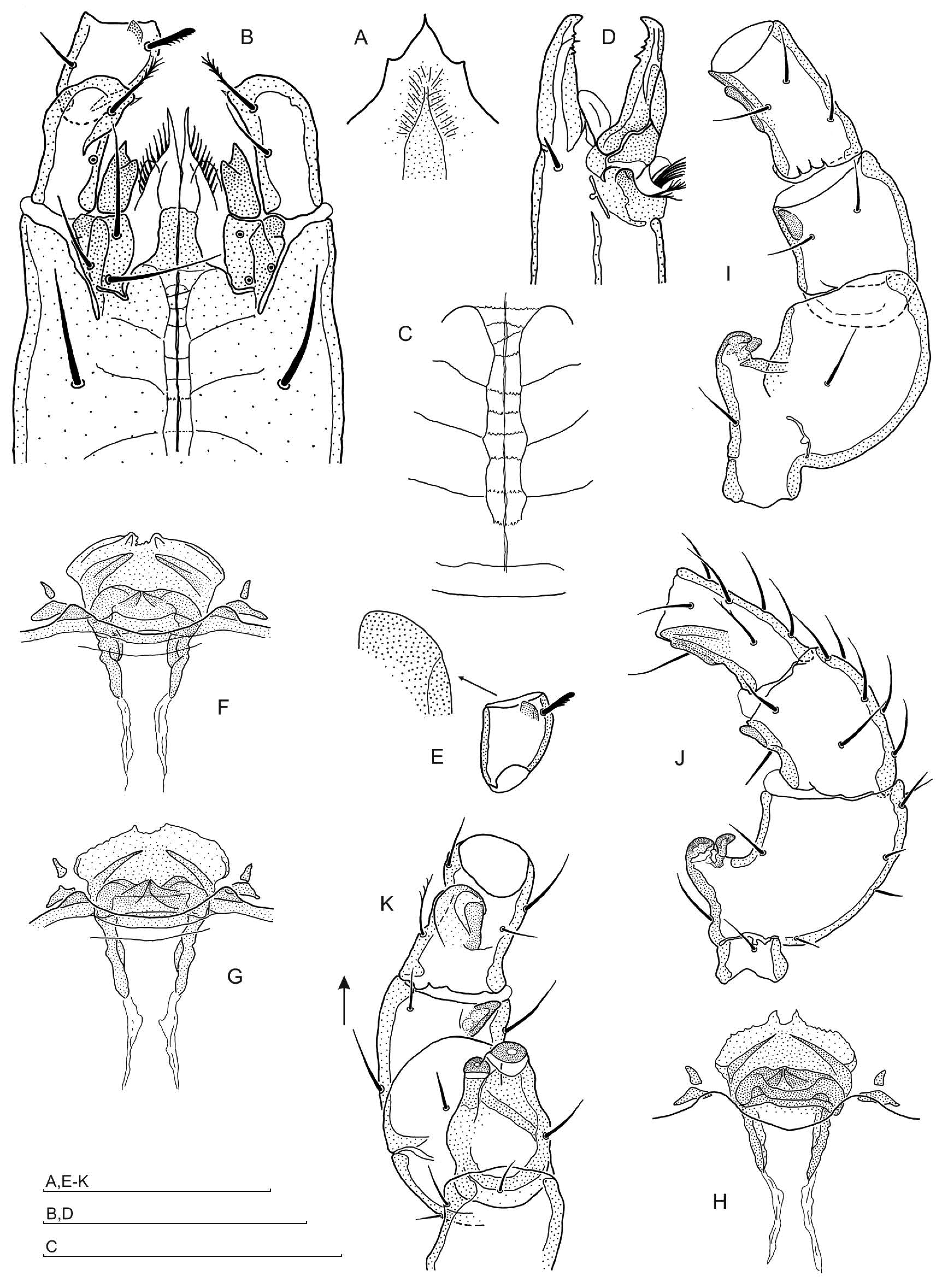
Diagnosis — In both sexes gland pores gv1 are present and pores gv2 are located on the surface of the flat cuticle; the gnathotectum is trispinate. In the female, the anterior wavy margin of the presternal plate is finely dentate; sternal shield has a fine granular band running axially; the endogynium is a narrow, ventro-dorsally oriented tube, visible in ventral perspective as a very small ring between the posterior paragynial lobes, sometimes open on one or both sides when the endogynial tube is obliquely oriented; the ventral margin of the endogynium in most specimens bears a triangular lamellar protrusion, located asymmetrically; ventrally, the endogynium is covered by a delicate endogynial lamella, dentate at the anterior margin. In the male, the corniculi have a prominent dent in the distal half; the hypostome has a narrow thickening ending anteriorly at the level of the corniculi bases; the pedipalpal femur bears a rectangular tubercle located anteroventrally, close to the anterolateral seta; the main spur on the femur II ends at the same level as the axillary process; spurs on genu II and tibia II are different, that on genu II is conical and close to the distal margin of the segment, that on tibia II is broad, low in the lateral perspective, and ending before the distal margin of the segment; the spur on tibia II has a margin shorter than its basal part.
Description
Female — Idiosoma brownish, 450 – 505 x 690 – 730 μm (width range x length range) (n=3). Length of podonotal setae: 39 – 44 μm (j1), 41 – 52 μm (r3), others 25 – 44 μm; opisthonotal setae very short, 9 – 12 μm; length of peritrematal groove 196 – 209 μm.
Gnathosoma — Gnathotectum trispinate (Fig. 1A), corniculi conical. Hypognathal groove narrow, with ca. ten very indistinct rows of denticles. Palpcoxal setae finely pectinate, anterior hypostomal seta h1 finely barbed, h2, h3 simple. Chelicera (Fig. 1B). Fixed digit with 2-3 denticles in front and two behind pilus dentilis, movable digit with three teeth. Pedipalp trochanter with seta v1 simple and v2 barbed distally, setation of pedipalp femur and genu unremarkable: seta al on pedipalp femur with pectinate posterior margin, setae al1 and al2 on genu spatulate, other setae simple.
Ventral idiosoma — Presternal plate (Fig. 1C) with a wavy and dentate anterior margin, posteriorly partly covered by the anterior margin of the sternum. Lateral platelets triangular or rhombic and free. Sternum (Fig. 1D) reticulated in the anterior part up to the level of pores iv2, anteriorly more pigmented, with a granular band of cuticle running axially; pores gv1 in sternal shield near its posterior margin. Setae of sternogenital region: 38 – 44 μm (st1), 38 – 41 μm (st2), 29 – 33 μm (st3), 31 – 39 μm (st4).
Genital region — Paragynial shields (Fig. 1E) reticulated, posterolateral protrusion short; metagynial sclerite arcuate and narrow, thickening near coxa III not present. Epigynial shield (Fig. 1F) with central prong well developed, anterior subapical thickenings absent, posterior thickenings running parallel and continuing laterally to the concavities for coxa IV, lateral hyaline protrusions of subapical structure triangular, slightly extending beyond the central prong margins. Endogynium (Fig. 1G-N) tubular, in situ visible as a small ring (external/internal diameter 13 – 14 / 5 – 7 μm), its opening usually guarded by asymmetric triangular lamella (Fig. 1G,K,L). When the tube is obliquely oriented to the vision axis, the ring is open on one side (Fig. 1H). Dissected endogynium (Fig. 1N) tubular with closed end directed more or less dorsally. Entrance of endogynium located ventrally and covered by endogynial lamella. The anterior margin of endogynial lamella is irregularly dentate (Fig. 1G, J-N). Gland pores gv2 are located on the surface of the flat cuticle. Opisthogaster with eight pairs of ventral setae 21 – 42 μm long.


Legs — Leg structure and setation unremarkable.
Male — Idiosoma brownish, 380 x 597 μm in holotype, 365 – 385 x 585 – 640 (n=2). Length of podonotal setae: 26 – 30 μm (j1), 28 – 30 μm (r3), others 16 – 25 μm; opisthonotal setae short, 8 – 20 μm; length of peritrematal groove 175 – 186 μm.
Gnathosoma — Gnathotectum trispinate (Fig. 2A), corniculi with deep distal incision on the ventroaxial side forming a very prominent dent (Fig. 2B). Hypostome with narrow thickening ending anteriorly at the level of the corniculi bases; hypognathal groove (Fig. 2C) as in the female, with ten fine rows of denticles. Palpcoxal setae finely pectinate, hypostomal setae simple. Chelicera (Fig. 2D). Fixed digit with a group of 3-4 denticles near pilus dentilis; movable digit bears three teeth. Pedipalp trochanter with seta v1 finely pectinate and v2 barbed; pedipalpal femur bears rectangular tubercle located anteroventrally, close to the anterolateral seta (Fig. 2E); pedipalp femur and genu setation unremarkable.
Ventral idiosoma — Presternal platelets (Fig. 2F-H) with two segments, anterior fine and sometimes curved, and posterior roughly triangular. Genital lamina (Fig. 2F-H) distinct, located in a concavity of the anterior sternal margin, which is flanked by tubercles on both sides. Anterior edge of lamina hyaline and bilobate, with two straight wedge-shaped thickenings directed towards central concavity in the anterior lamellar margin, lateral projections rounded, subgenital microsclerite well sclerotised, with two tooth-like thickenings apically meeting at the axial line. Sternogenital shield with distinct regular reticulation; excipulum absent. Sternal setae length: 33 – 39 μm (st1), 27 – 29 μm (st2), 27 – 38 μm (st3), setae st4 26 – 30 μm, opisthogaster setae 22 – 37 μm.
Legs — Leg I, III, IV unremarkable. Leg II (Fig. 2I-K) as follows: coxa II with a ridge of 5-6 denticles anterolaterally, accompanied by a basal denticle; the main spur on the femur ending at the same level as the axillary process; spurs on genu II and tibia II different, that on genu II conical and located close to the distal margin of the segment, that on tibia II broad, low in the lateral perspective and ending before the distal margin of the segment; its margin shorter than the basis as a result of the distally located incision (Fig. 2J).

Etymology — The specific name refers to Calpe, the original Latin name of the Rock of Gibraltar, the type locality of the species.
Material examined — Holotype male (slide no. 2489), 16.10.2014, Gibraltar, 36°7.621'N, 5°20.919'W, alt. ca. 115 m a.s.l., dry leaf litter beside street; 6 female and 1 male paratypes (slides no. 2486, 2487, 2488, 2489, 2490, 2502), ibid.; 1 female and 2 male paratypes (slides no. 2475, 2476, 2477A-D), 15.10.2014, 36°8.596'N, 5°20.795'W, alt. ca. 212 m a.s.l., dry leaf litter under bushes; 1 male paratype (slide no. 2053), 10.01.1997, Gibraltar, Alameda Botanic Gardens, approx. coordinates 36°7.890'N, 5°21.055'W, alt. ca. 37 m a.s.l., leaf litter under trees, leg. Prof. W. Niedbala, Adam Mickiewicz University, Poznan, Poland. Types are deposited in the Zoological Museum of the Jagiellonian University, Cracow, Poland.
Remarks — Holoparasitus calpetanus n. sp. belongs to the Holoparasitus mallorcae species-group, which is characterised as follows (Juvara-Bals 2008). In females, (1) presternal plate bears denticles or granules, especially at the anterior margin, lateral platelets free; (2) sternum with an axial reticulated or granular band; (3) the thickening of the anterior paragynial edge facing coxa III absent; (4) fixed cheliceral digit with five denticles; (5) gv2 in unmodified flat cuticle. The first two character states are usually present together, but in a few species only one of them is present. In males, (1) hypostome with the tongue-shaped, sclerotised central part, extended in many species antero-laterally between the corniculi; (2) hypostomatic setae on a piece of cuticle separated by incisions of soft cuticle; (3) sternogenital shield without an excipulum; (4) leg II: coxa II possesses anterolaterally denticulate ridge and a basal denticle, femoral main spur and axillary process thumb-like and short.
Holoparasitus calpetanus is included in the Holoparasitus mallorcae species-group since both females and males show all species-group character states. On the other hand, the female of H. calpetanus differs from other species belonging to the species-group by the following characteristics: in H. algiersensis Juvara-Bals, 2008 and H. eivissa Juvara-Bals, 2008 gv1 glands are absent. Holoparasitus calpetanus differs from H. siculus (Berlese, 1906), as the central prong of the epigynium in this species is evidently elongate. In contrast to all other species of the H. mallorcae species-group, the endogynium in H. calpetanus is tubular and narrow, whereas in the remaining species it is large and saccular. In the male, H. calpetanus differs from H. algiersensis Juvara-Bals, 2008, H. eivissa Juvara-Bals, 2008 and H. singularis Juvara-Bals, 2008 since it possesses gv1 glands. Compared to other species of this species-group H. calpetanus is most similar to H. siculus (Berlese, 1906), H. mahnerti Juvara-Bals, 2008, H. mallorcae Juvara-Bals, 1975 and H. variabilis Juvara-Bals, 2008. It differs from H. siculus (Berlese, 1906) by a gnathotectum with an acute central prong and regular lateral prongs, whereas from H. mahnerti Juvara-Bals, 2008 by the absence of a basal protrusion on the axillary process of femur II. Corniculi with prominent dents on their distal halves distinguish H. calpetanus from H. mallorcae Juvara-Bals, 1975 and H. variabilis Juvara-Bals, 2008, both showing corniculi with medium-sized dents on proximal halves.
ZOOBANK: 5326BC8D-B3D4-450C-B686-7F7B4F836AA0 ![]()
Diagnosis — In both sexes gland pores gv1 are present, and pores gv2 are located in unmodified flat cuticle. In the female, the gnathotectum is trispinate, presternal plate is ribbon-shaped, only slightly narrowed medially, with obliquely cut ends, no denticles or corrugation are present; lateral platelets free and wedge-shaped; sternal plate reticulation pronounced, axial granular or reticulated band absent, anterior sternal margin concave between st1 setae and frequently with small concavities laterally to sternal pores iv1; posterior paragynial lobes in close proximity, metagynial sclerite heavily sclerotised and located close to adaxial edge of paragynium, its antiaxial margin straight whereas the adaxial margin forms a rounded protrusion matching the concavities in the anterior subapical thickening of the epigynium; epigynial central prong much wider than the subapical structure, which is narrow and anteriorly tripartite, with lateral hyaline protrusions rounded and extending slightly beyond the central prong margins; endogynium very small in ventral perspective, with heavily sclerotised roundish structure located centrally, and the lateral parts bearing minute denticles; anteriorly, endogynium forming a lamellar protrusion. In the male, the gnathotectum the solid central prong is rounded apically, lateral prongs absent; corniculi conical; hypostome normal; hypognathal groove clearly visible; chelicera fixed digit straight and edentate with a characteristic lamellar protrusion laterally, movable digit solid and curved adaxially, bearing one dent; pilus dentilis minute, settled in orifice in lateral protrusion of fixed digit; palpcoxal setae scarcely pectinate and hypostomatics simple; palp trochanter with stout simple seta v1 and longer seta v2 barbed terminally, both setae located in close proximity and separated by a prominence; palp femur with ventral tubercle near the anterolateral seta; sternal shield with a regular reticulation except the roundish area between coxa II and coxa III, where the reticulation is weak and the cuticle porous; genital lamina rounded, located in a concavity of the sternal margin, and flanked by the sclerotised prominences; apical segment of femur II main spur circular in the ventral perspective, axillar process wedge-like, spur on genu II moderate, the one on tibia II – low, roundish and broad; genu II extended anterolaterally in the distal part.
Description
Female — Idiosoma brownish, 475-495 x 670-675 μm (width range x length range) (n=5). Length of podonotal setae: 26-30 μm (j1), 25-26 μm (r3), others 14-17 μm; opisthonotal setae very short, 8-12 μm; length of peritrematal groove 209-215 μm.
Gnathosoma — Gnathotectum trispinate (Fig. 3A), corniculi conical. Hypognathal groove narrow, with ca. ten weakly pronounced rows of denticles. Palpcoxal and hypostomatic setae simple. Chelicera (Fig. 3B). Fixed digit with two denticles in front of pilus dentilis, one larger and lamellar, covering pilus dentilis, and two small denticles behind pilus dentilis; the latter are located somewhat below the digit edge, which is lamellar and arcuate. The movable digit bears three teeth, pedipalp trochanter with seta v1 simple, v2 longer and barbed distally; femur and genu setation unremarkable.
Ventral idiosoma — Presternal plate (Fig. 3C) ribbon-shaped with obliquely cut ends and only slightly narrowed medially, and with a sinuous anterior margin, no denticles or corrugation present. Lateral platelets are free and wedge-shaped. Sternal shield (Fig. 3D) anterior margin with shallow concavity between st1 setae and usually with small concavities laterally to iv1 sternal pores, reticulation well developed. Pores gv1 on sternal shield near posterior sternal margin. Setae of sternogenital region: 39 – 46 μm (st1), 50 – 59 μm (st2), 44 – 51 μm (st3), 33 – 39 μm (st4).
Genital region — Paragynial shields (Fig. 3E) reticulated, posterolateral protrusions well developed, roundish or sometimes asymmetrical. Posterior paragynial lobes in close proximity. Adaxial paragynial edge thickened and metagynial sclerite heavily sclerotised, located close to the adaxial paragynial edge; antiaxial margin of metagynial sclerite straight or slightly curved, adaxial margin forming a rounded protrusion matching concavity in the anterior subapical thickening of the epigynium (Fig. 3E). Thickening facing coxa III is absent. Central prong of epigynium (Fig. 3F,G) with convex margins, wider than the subapical structure, which is narrow and anteriorly tripartite. Lateral hyaline protrusions are rounded and extend only slightly beyond the margins of the central prong. Endogynium (Fig. 3H,I) is small, with heavily sclerotised roundish structure located centrally and with the lateral, less sclerotised parts bearing minute denticles; anteriorly, endogynium forms a lamellar tongue-like protrusion directed anteriorly and frequently indented apically. Gland pores gv2 in unmodified flat cuticle. Opisthogaster with eight pairs of ventral setae 15 – 42 μm long.


Legs — Leg structure and setation unremarkable.
Male — Idiosoma brownish, 387 – 425 x 580 – 640 (n=5). Length of podonotal setae: 26 – 29 μm (j1), 16 – 18 μm (r3), others 9 – 14 μm; opisthonotal setae short, 10 – 12 μm; length of peritrematal groove 199 – 207 μm.
Gnathosoma — Gnathotectum with a solid central prong rounded apically, lateral prongs absent, gnathotectum margins convex (Fig. 4A). Corniculi (Fig. 4B) conical. Hypostome normal, hypognathal groove rather wide, and ca. 10 rows of denticles moderately visible. Palpcoxal setae scarcely pectinate, hypostomal setae simple. Chelicera (Fig. 4C-F). Fixed digit straight and edentate with a characteristic lateral lamellar protrusion showing a small orifice at the base of minute pilus dentilis (Fig. 4D,F). The protrusion is clearly visible in the ventral perspective (Fig. 4D). Movable digit solid and curved adaxially; bearing one tooth, in some specimens followed distally by 1-2 minute denticles. Pedipalp trochanter (Fig. 4B,G) with stout seta v1 and longer seta v2 barbed terminally, both setae located in close proximity and separated by a small prominence. Pedipalp femur (Fig. 4B) with ventral tubercle near the anterolateral seta; both femur and genu setation unremarkable.
Ventral idiosoma — Presternal platelets small and roughly triangular (Fig. 5A). Genital lamina (Fig. 5B) roundish with sclerotised margin and heavily sclerotised subgenital microsclerite. The lamina is located in the concavity of the anterior sternal margin, flanked by the sclerotised tubercles on both sides of the concavity. Reticulation of the sternogenital shield (Fig. 5A) clearly visible laterally, but in central part between coxa II and coxa III weakly pronounced, and the sternal cuticle is porous. Excipulum absent. Sternal setae length: 34 – 39 μm (st1), 30 – 33 μm (st2), 31 – 35 μm (st3), setae st4 27 – 30 μm, opisthogastral setae 21 – 37 μm.

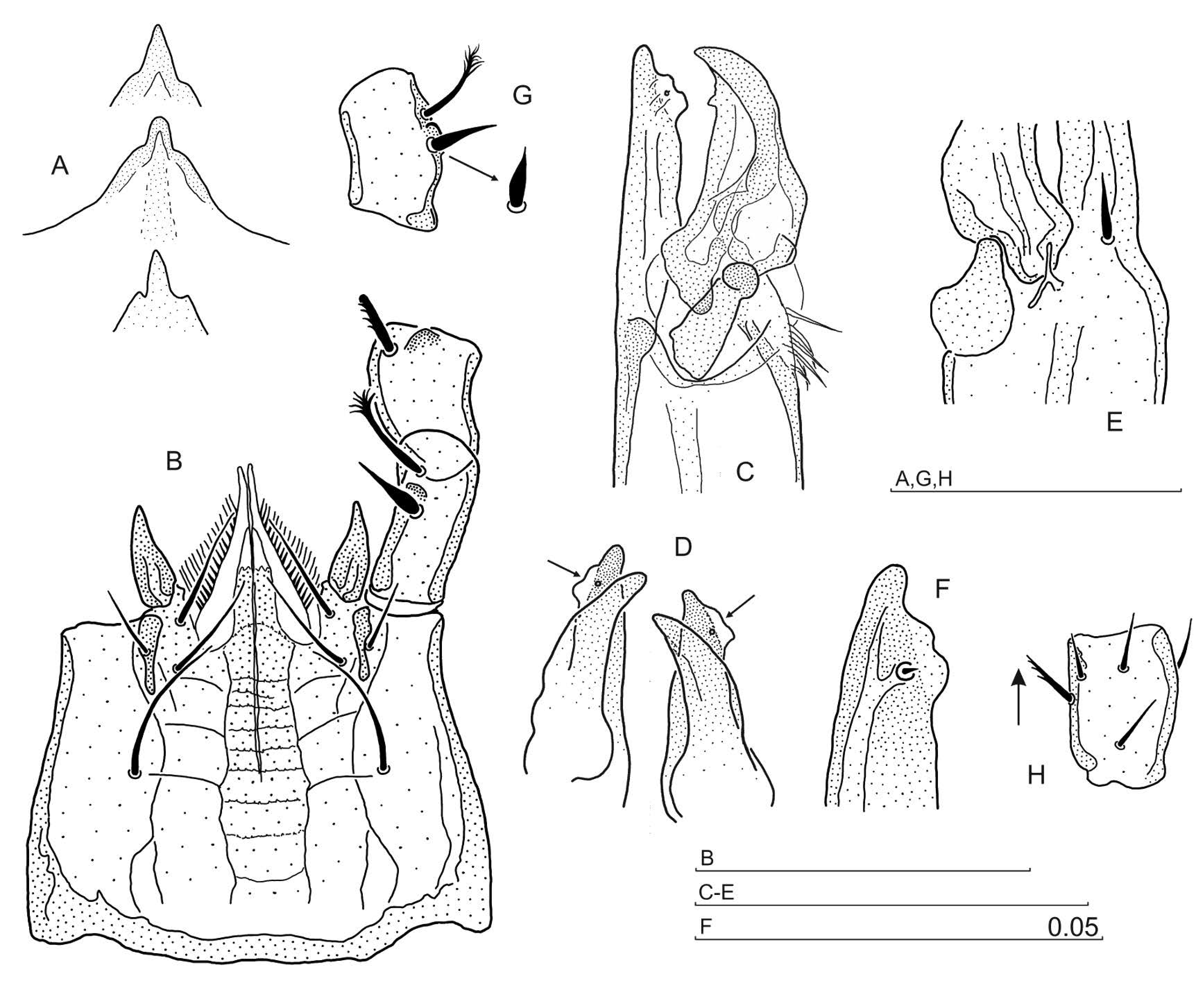
Legs — Leg I, III, IV largely unremarkable, with simple setae, but anterolateral setae on trochanter I and trochanter II, which are thicker and pectinate distally (Fig. 4H). Leg II (Fig. 5C-E) spurred as follows: apical segment of femur II main spur circular in the ventral perspective, whereas the axillar process wedge-like; spur on genu II moderate, that on tibia II low, roundish and broad. genu II is extended anterolaterally in the distal part (Fig. 5D,E).


Etymology — The specific name refers to its type locality, Fanes in Val Travenanzes, Dolomites, Italy.
Material examined — Holotype female (slide no. 1620), 18.08.2001, Cortina d'Ampezzo, Dolomites, Italy, 46°34.329'N, 12°6.789'E, alt. ca. 1311 m a.s.l., moss on tree stump in a spruce forest near the road to Olympia Camping; 8 female and 12 male paratypes (slides no. 2000, 2001, 2002), 12.09.2005, Val Travenanzes, Dolomites, close to Fanes waterfalls (Cascate di Fanes), moss and litter in a spruce forest, 46°35.723'N, 12°5.185'E, alt. ca. 1455 m a.s.l.; 4 female and 4 male paratypes (slides no. 2162, 2163), 7.09. 2007, Val Travenanzes, Dolomites, litter and moss in a spruce forest, 46°35.127'N, 12°4.605'E, alt. 1635 m a.s.l.
Other material: 3 females, 4 males (slides no. 1640, 1641, 1713-1717), 10.09.2002, Cortina d'Ampezzo, Dolomites, near Olympia Camping, litter in a spruce forest, 46°34.329'N, 12°6.789'E, alt. ca. 1311 m a.s.l.; 1 male (slide no. 2011), 13.09.2005, near Olympia Camping, Cortina d'Ampezzo, Dolomites, thick layer of moss covering soil and logs in a spruce forest, 46°34.335'N, 12°6.746'E, alt. ca. 1320 m a.s.l.; 13 females, 24 males (slides no. 2179, 2180, 2182), 30.09.2008, near Dobbiaco, Dolomites, moss under trees, 46°41.520'N, 12°12.450'E, alt. ca. 1626 m a.s.l. Types are deposited in the Zoological Museum of the Jagiellonian University, Cracow, Poland, whereas the other material remains in the Author's collection.
Holoparasitus crassisetosus species-group, newly defined. Species included:
Holoparasitus crassisetosus Juvara-Bals and Witalinski, 2000
Holoparasitus digitiformis Juvara-Bals and Witalinski, 2000
Diagnosis of the species group — In both sexes (1) gnathotectum trispinate; (2) gland pores gv1 present; (3) gland pores gv2 located in unmodified flat cuticle. Female — (1) gnathotectum regularly trispinate; (2) presternal plate with the anterior margin smooth, lateral platelets free, or one-sidedly accreted to presternal plate; (3) sternum entire, with regular reticulation, axial granular or reticulated band absent; (4) sternal anterior margin developed normally; (5) posterior paragynial lobes in close proximity to each other; (6) the thickening of the anterior paragynial edge facing coxa III present; (7) central epigynial prong moderate and triangular, the thickening of subapical epigynial structure well visible, compact, anteriorly rounded; (8) well sclerotised endogynium very small and roundish, with a protrusion directed dorsally. Male — (1) gnathotectum trispinate-type, but the central prong well pronounced and acute, whereas the lateral prongs minute; (2) hypostome regularly triangular and moderately sclerotised; (3) hypostomatic setae on entire cuticle, but not on the piece of cuticle separated by lateral and posterior soft-cuticular incisions; (4) corniculi with a circular lamellar protrusion located adaxially; (5) excipulum on the sternum absent; (6) genital lamina with a well sclerotised subgenital microsclerite and long, acute hyaline lateral protrusions; (7) pedipalp trochanter with seta v1 simple and thickened basally, seta v2 barbed terminally; (8) pedipalp femur bears a ventral tubercle; (9) femur II spur conical, with a small terminal segment, axillary process settled on the slope of femoral spur; (10) spur on genu II small and semicircular, located in the middle of the segment.
Distribution — Both species of this group are found in the central part of Italy, Tuscany, whereas H. digitiformis is also found in Sardinia (Juvara-Bals and Witalinski 2000).
Remarks — The new species H. fanes is most similar to the H. crassisetosus species-group. It shows all the character states shared by both sexes (1-3). The females of H. fanes possess some characteristics of the group (1-5), but details of the genital orifice area are different. The males of H. fanes are similar to the H. crassisetosus group in gnathotectum and hypostome structure (1-3), sternum (5) and pedipalp (7, 8), but corniculi (4), genital lamina (6) and leg II structure (9, 10) are different.
The female of H. fanes can be distinguished from the H. crassisetosus species-group by the anteriorly tripartite, rather than rounded, thickening of the subapical epigynial structure. In the male, a porous area in the sternum center is present only in H. fanes. Moreover, the corniculi are conical and tibia II regularly spurred. These characteristics differentiate H. fanes from H. inornatus (Berlese, 1906) and H. intermedius (Holzmann, 1969), both species with indented corniculi. H. megacalcaratus Schmölzer, 1995 possesses conical corniculi, but with only a single, large main spur on femur II.
The author would like to express his gratitude to Professor W. Niedbala, Adam Mickiewicz University, Poznan, who kindly made available the material collected by himself on Gibraltar in 1997, comprising the H. calpetanus male, for the purpose of the present study.
Berlese A. 1906 — Monografia del genere Gamasus Latr. — Redia, 3: 66-304.
Holzmann C. 1969 — Die Familie Parasitidae Oudemans, 1901 — Acarologie, 13: 3-55 + 23 Plates.
Hyatt K.H. 1987 — Mites of the genus Holoparasitus Oudemans, 1936 (Mesostigmata: Parasitidae) in the British Isles — Bull. Br. Mus. nat. Hist. (Zool.), 52: 139-164. doi:10.5962/bhl.part.18305 ![]()
Juvara-Bals I. 1975 — Sur le genre Holoparasitus Oudemans, 1936 et sur certains caractères morphologiques de la famille Parasitidae Oudem. (Parasitiformes) — Acarologia, 17: 384-409.
Juvara-Bals I. 2008 — New species of Holoparasitus Oudemans, 1936 (Acari, Parasitidae) from Spain, North Africa and Madeira Islands — Revue suisse Zool., 115: 37-84. doi:10.5962/bhl.part.80420 ![]()
Juvara-Bals I., Witalinski W. 2000 — Description of five new species of Holoparasitus s. str. with redescription of H. apenninorum (Berlese, 1906) and H. cultriger (Berlese, 1906) from Italy and Spain (Acari, Gamasida, Parasitidae) — Revue suisse Zool., 107: 3-30. doi:10.5962/bhl.part.80116 ![]()
Juvara-Bals I., Witalinski W. 2006 — Two new species of the genus Holoparasitus Oudemans from the Mediterranean Basin – Algeria and Sardinia (Acari: Gamasida: Parasitidae) — Genus, 17: 437-448.
Micherdzinski W. 1969 — Die Familie Parasitidae Oudemans, 1901 (Acarina, Mesostigmata) — Panstwowe Wydawnictwo Naukowe, Kraków, 690 pp.
Oudemans A.C. 1936 — Kritisch Historisch Overzicht der Acarologie. III Ged. 1805-1850, Band A. E. J. Brill, Leyden, XVI + 430 pp.
Schmölzer K. 1995 — Landmilben aus den östlichen Karawanken (Acarina, Parasitiformes) — Carinthia II, 185: 499-512.
Witalinski W. 2004 — Holoparasitus excipuliger (Berlese, 1906) in Hungary: a second world locality and redescription (Acari: Gamasida: Parasitidae) — Genus, 15: 425-434.
Witalinski W. 2006 — New mites of the genus Holoparasitus Oudemans, 1936 from northern Austria and Karavanke (Acari: Parasitidae) — Zootaxa, 1320: 15-27.
Witalinski W., Skorupski M. 2002 — Genus Holoparasitus Oudemans, 1936 in Berlese Acaroteca (Acari: Gamasida: Parasitidae). Part I — Redia, 85: 37-60.
Witalinski W., Skorupski M. 2003a — Genus Holoparasitus Oudemans, 1936 in Berlese Acaroteca (Acari: Gamasida: Parasitidae). Part II — Redia, 86: 17-22.
Witalinski W., Skorupski M. 2003b — A new species of Holoparasitus Oudemans, 1936 from North Italy (Acari: Gamasida: Parasitidae) — Genus, 14: 431-438.
Witalinski W., Skorupski M. 2007 — Two new species of Holoparasitus Oudemans, 1936 from Italy – Tuscany and Trentino (Acari: Gamasida: Parasitidae) — Genus, 18: 125-138.
Descriptions lacking illustrations of the body dorsum, female subcapitulum, female ventrianal shield, and legs are hardly useful. And to indicate “Leg structure and setation unremarkable” (except for male leg II) is inadequate.


2016-08-02
Date accepted:
2016-09-28
Date published:
2016-12-16
Edited by:
Faraji, Farid

This work is licensed under a Creative Commons Attribution 4.0 International License
2017 Witalinski, Wojciech
 Download article
Download the citation
RIS with abstract
(Zotero, Endnote, Reference Manager, ProCite, RefWorks, Mendeley)
RIS without abstract
BIB
(Zotero, BibTeX)
TXT
(PubMed, Txt)