


Two new species of Diptilomiopus Nalepa (Acari: Eriophyoidea) from India
Chakrabarti, Samiran1 ; Sur, Surajit2 and Sarkar, Sanjay3
1✉ Post-Graduate Department of Zoology, Vidyasagar College, 39, Sankar Ghosh Lane, Kolkata-700006, India.
2Post-Graduate Department of Zoology, Vidyasagar College, 39, Sankar Ghosh Lane, Kolkata-700006, India.
3Post-Graduate Department of Zoology, Vidyasagar College, 39, Sankar Ghosh Lane, Kolkata-700006, India.
2019 - Volume: 59 Issue: 3 pages: 383-394
https://doi.org/10.24349/acarologia/20194337ZooBank LSID: 465F8BB9-9D65-4138-B433-D0D12331EF04
Original research
Keywords
Abstract
Introduction
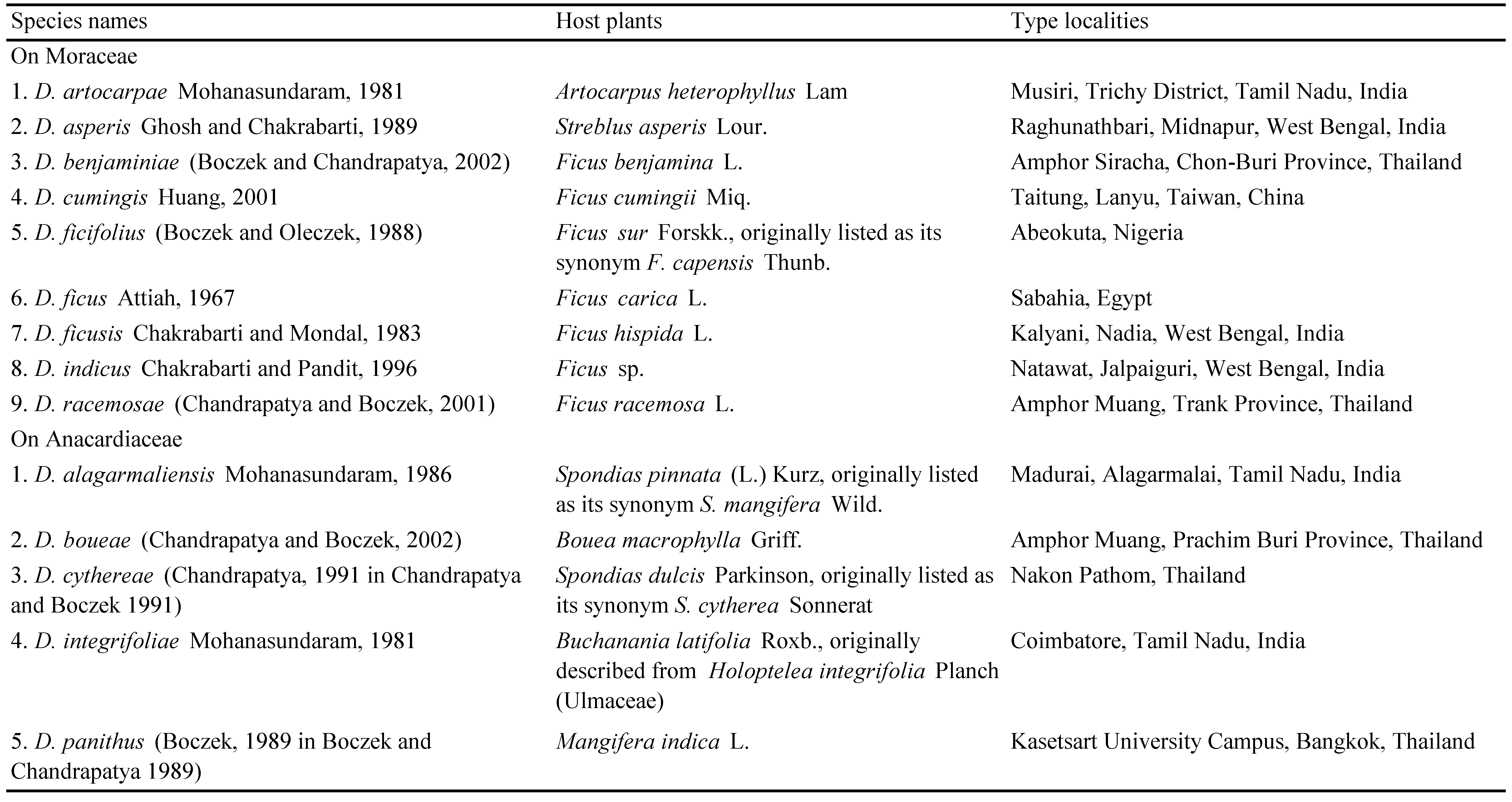
During periodical samplings of eriophyoid mites from different host plants in West Bengal, India, two Diptilomiopus species on dye fig, Ficus tinctoria subsp. gibbosa (Blume) Corner (Moraceae) and mango, Mangifera indica L. (Anacardiaceae) were collected. In general, mites in the Diptilomiopus Nalepa (1916) are mostly distributed in the Oriental region. Newkirk and Keifer (1975) set Sectipes Keifer (1962) as a junior synonym of Diptilomiopus. Similarly, Hong and Zhang (1997) set Vilaia Chandrapatya and Boczek (1991) as a junior synonym of Diptilomiopus. Craemer et al. (2005) confirmed this synonymy and transferred all additional 16 species described in Vilaia to Diptilomiopus. At present, 102 valid species of Diptilomiopus are known (Craemer et al., 2017; Sur et al., 2018; Amrine 2019, personal communication). So far 9 species of Diptilomiopus have been described on plants of the family Moraceae and 5 species on Anacardiaceae (Table 1). In this account, descriptions of two further Diptilomiopus species and keys for separating these from closely related species are provided. A note has been provided on Diptilomiopus strebli (Boczek, 1992 in Boczek and Chandrapatya 1992) infesting Streblus asperis Lour. (Type locality- Dusit zoo, Bangkok, Thailand) and Diptilomiopus holoptelus Chakrabarti and Mondal, 1983 infesting Holoptelea integrifolia Planch (Type locality- Kalyani, Nadia, India) suspecting their possible synonymies with D. asperis Ghosh and Chakrabarti, 1989 and D. integrifoliae Mohanasundaram, 1981, respectively.

Material and methods
Eriophyoid mites were collected and mounted as described by Chakrabarti et al. (2017) and Hoyer's medium was used for mounting the specimens. The terminology and classification given by Lindquist (1996) and Amrine et al. (2003), respectively, are followed here. The specimens were examined with a phase contrast microscope Leica DM3000 and photographs were taken with Leica DFC295 camera. All measurements were made following Amrine and Manson (1996) and de Lillo et al. (2010) and are given in micrometres (µm). Measurements and means are rounded off to the nearest integer and refer to the length of the morphological characters unless specified otherwise. Drawings were made following de Lillo et al. (2010) and Amrine et al. (2003). In the text, measurements of the holotype are followed by the range of measurements of the paratypes plus holotype given in parentheses. Original descriptions and measurements of D. strebli (Boczek, 1992 in Boczek and Chandrapatya 1992) and D. holoptelus Chakrabarti and Mondal, 1983 were compared with D. asperis Ghosh and Chakrabarti, 1989 and D. integrifoliae Mohanasundaram, 1981 suspecting their possible synonymies. All type specimens are now deposited in the collection of the Post-Graduate Department of Zoology, Vidyasagar College, Kolkata- 700006, India. After publication, holotypes and paratypes will be deposited in public institutions: one slide with paratypes of each species will be deposited in the National Pusa Collection, Indian Agricultural Research Institute, New Delhi; the holotypes and the remnant paratypes will be deposited in the National Zoological Collection, Zoological Survey of India, Kolkata.
Results
Family: Diptilomiopidae Keifer, 1944
Subfamily: Diptilomiopinae Keifer, 1944
Diptilomiopus indogangeticus n. sp.
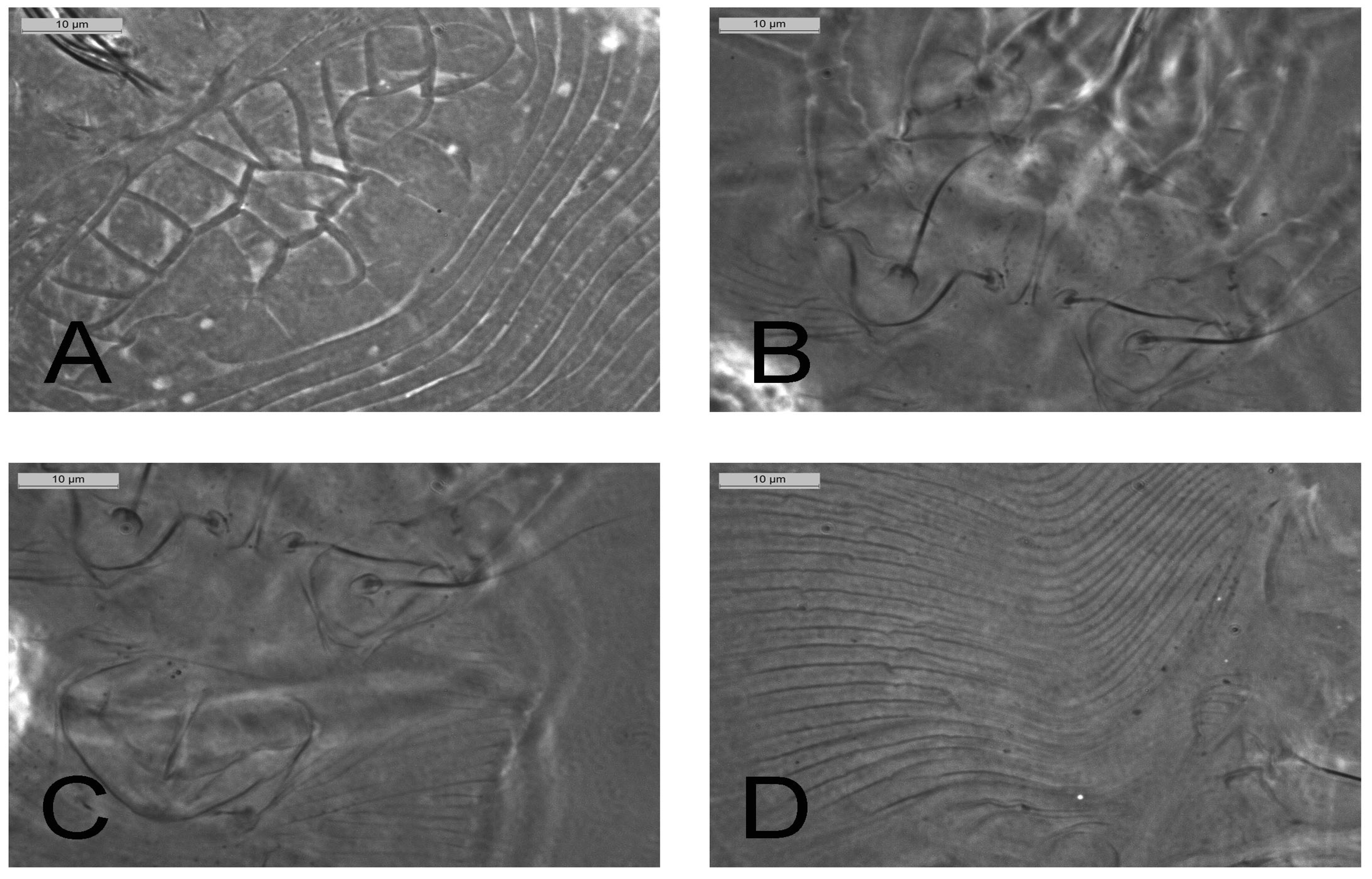
(Figures 1, 2)
Diptilomiopus ficivorus Sarkar, 2011: 120. Invalid name, a thesis name.
ZOOBANK: 5F402A24-ED4A-49FF-AFDD-7AB6C7CB6878 ![]()
Description


Female (n=15) — Live colour brown. Body fusiform, 145 (140–151), 67 (66–67) wide. Gnathosoma: 39 (38–39), abruptly curved down, palp setae d 6 (5–6), palp setae ep 3 (2–3). Prodorsal shield: 22 (22–24), 44 (44–46) wide, with complete network of cells composed of one central cell and a row of 12 cells on the anterior margin, a row of 7 cells on the posterior margin and in between these two rows another 6 cells; scapular tubercles 2 (2–3) and located ahead of shield margin; scapular setae sc absent. Leg I: 23 (23–24), femur 17 (16–17), femoral setae bv absent; genu absent; tibia 6 (5–6), tibial setae l′ absent; tarsus 6 (5–6), tarsal setae ft′ 30 (29–30), tarsal setae ft″ 29 (29–31), setae u′ 2 (2–3); empodium em 10 (10–11), deeply divided, each branch 6-rayed, solenidion ω 9 (9–10), knobbed. Leg II: 21 (20–21), femur 13 (13–14), femoral setae bv absent; genu absent; tibia 4 (4–5), tibial setae l′ absent; tarsus 5 (5–6), tarsal setae ft′ 25 (25–27), tarsal setae ft″ absent, setae u′ 2 (2–3); empodium em 10 (10–11), deeply divided, each branch 6-rayed, solenidion ω 9 (9–10), knobbed. Coxae I: 14 (14–15), with granules, sternal line present; setae 1b absent; setae 1a 21 (21–22) and 8 (8–9) apart; coxae II: 14 (14–15), with granules as in coxae I, setae 2a 31 (30–31) and 15 (15–16) apart, setae 1a located little ahead of 2a. Opisthosoma: Dorsal annuli 64 (64–66) with two pairs of lateral ridges; ventral annuli 62 (60–63), with small, roundish microtubercles, last 8 (7–8) ventral annuli with elongated and linear microtubercles; setae c2 absent; setae d 12 (10–12) on ventral annulus 20; setae e 15 (14–15) on ventral annulus 32; setae f 19 (19–21) on ventral annulus 49; setae h1 absent, setae h2 32 (30–32). Genital coverflap: 16 (16–17), 22 (22–23) wide, with granules present on the basal part, setae 3a 7 (7–8). Internal genitalia: Apodeme short, spermathecae rounded with short funnel-like spermathecal tubes.
Male — Not found.
Etymology — The specific epithet is masculine in gender and `indogangeticus' is derived from the locality of collections of infesting plants occurring Indo-Gangetic plane on the southern side of the River Ganges in Murshidabad district, West Bengal.
Type material — Holotype (circled): female (slide no. 1825/98/2016), 07 Aug. 2016, Chunakhali (24˚07′44″N, 88˚17′40″E, alt. 20 m a.s.l.), Murshidabad, West Bengal, India, from Ficus tinctoria subsp. gibbosa (Blume) Corner (Moraceae), coll. S. Sur. Paratypes: 2 females and 4 nymphs in the slide bearing the holotype and 15 females (slide no. 1826-1828/98/2016), collection data as in the holotype.
Additional specimens: 7 females and 9 nymphs (slide no. 1392/72/2006), 14 Oct. 2006, Amriti (24˚20′38″N, 87˚05′29″E, alt. 30 m a.s.l.), Malda, West Bengal from the same host plant, coll. S. Sarkar.
Relation to the host plant — Mites are vagrant on lower surface of leaves showing no apparent damage symptoms.
Differential diagnosis — Diptilomiopus indogangeticus n. sp. shows many similarities with D. cayratus Cheng et al., 2012; D. euscaphiae Wang et al., 2009; D. lithocarpi Wang et al., 2009; D. ligustri Wang et al., 2009 and D. terstroemiae Wang et al., 2009 in having reticulated cellular network on prodorsal shield along with scapular tubercles and presence of palp setae d and ep. However, D. indogangeticus n. sp. can be separated from the above mentioned species by the absence of h1 setae and having 6-rayed empodium. The above Diptilomiopus species are very close and can be separated by the following key.
Remarks — The name Diptilomiopus ficivorus appeared previously in the Ph.D thesis by Sarkar (2011) for this species.
Key to the closely related species (mentioned in the differential diagnosis) of Diptilomiopus indogangeticus
1. Empodium 3-rayed; on Cayratia japonica (Thunb.) Gagnep (Vitaceae)
...... D. cayratus Cheng et al., 2012
— Empodium at least 6-rayed
...... 2
2. h1 setae absent; palp setae v lacking; empodium 6-rayed; on Ficus tinctoria subsp. gibbosa (Blume) Corner (Moraceae)
...... D. indogangeticus n. sp.
— h1 setae present; palp setae v present; empodium 7-rayed
...... 3
3. Ventral annuli 62–64; genital coverflap sculptured with only granules; on Lithocarpus glaber (Thunb.) Nakai (Fagaceae)
...... D. lithocarpi Wang et al., 2009
— Ventral annuli at least 77; genital coverflap either with granules and small lines or only sculptured with basal short lines
...... 4
4. Genital coverflap sculptured with basal short lines, dorsal annuli 60–71; on Ligustrum quihoui Carr. (Oleaceae)
...... D. ligustri Wang et al., 2009
— Genital coverflap with granules either at base or on whole coverflap region; dorsal annuli either 49–54 or 56–62
...... 5
5. Prodorsal shield cells arranged in 4 rows and without unpaired central cell; dorsal annuli 49–54; granules present only at basal part of genital coverflap; on Ternstroemia nitida Merr. (Theaceae)
...... D. terstroemiae Wang et al., 2009
\cledetermination{— Prodorsal shield cells arranged in 3 rows and with an unpaired central cell; dorsal annuli 56–62; granules present on whole genital coverflap; on Euscaphis japonica (Thunb.) Kanitz (Staphyleaceae)} {D. euscaphiae Wang et al., 2009}
A further key can separate the Diptilomiopus species associated with plants within the family Moraceae.
\texorpdfstring{Key to the Diptilomiopus associated with Moraceae}{Key to the Diptilomiopus associated with Moraceae}
1. Genital coverflap with longitudinal scorings
...... 2
— Genital coverflap either granulated or smooth
...... 3
2. Prodorsal shield with median line present on both anterior and posterior 0.25 parts; 7-rayed empodium; on Ficus hispida (L.)
...... D. ficusis Chakrabarti and Mondal, 1983
— Prodorsal shield without median line; 5-rayed empodium; on Artocarpus heterophyllus Lam.
...... D. artocarpae Mohanasundaram, 1981
3. Accessory setae h1 present; on Ficus benjamina L.
...... D. benjaminiae (Boczek and Chandrapatya, 2002)
— Accessory setae h1 absent
...... 4
4. Smooth genital coverflap; both coxae smooth; on Streblus asperis Lour.
...... D. asperis Ghosh and Chakrabarti, 1989
— Granulated genital coverflap; at least one coxa ornamented
...... 5
5. Both coxae granulated; prodorsal shield with 25 cells; on Ficus tinctoria subsp. gibbosa (Blume) Corner
...... D. indogangeticus n sp.
— Either both coxae or at least one coxae smooth; prodorsal shield with less than 25 cells
...... 6
6. Empodium 4-rayed; on Ficus carica (L.)
...... D. ficus Attiah, 1967
— Empodium at least 6-rayed
...... 7
7. Opisthosoma with smooth dorsal and ventral annuli; on Ficus cumingii (L.)
...... D. cumingis Huang, 2001
— Opisthosoma with smooth dorsal and either microtuberculated or microstriated ventral annuli
...... 8
8. Ventral annuli with microstriations; 22 cells on prodorsal shield; on Ficus racemosa L.
...... D. racemosae (Chandrapatya and Boczek, 2001)
— Ventral annuli with microtubercles; either 26 or 18 cells on prodorsal shield
...... 9
9. 26 cells on prodorsal shield with a central cell; both coxae smooth; on Ficus sp.
...... D. indicus Chakrabarti and Pandit, 1996
— 18 cells on prodorsal shield without central cell; only coxae II smooth; on Ficus capensis Thunb.
...... D. ficifolius} (Boczek and Oleczek, 1988)
Diptilomiopus mohanasundarami n. sp.
(Figures 3, 4)
Diptilomiopus mangiferae Sarkar, 2011: 125. Invalid name, a thesis name.
ZOOBANK: http://zoobank.org/act/5FCE349F-A4EE-4EC0-9D12-49A3CAFB2DA7 ![]()
Description

Female (n=15) — Live colour brown. Body fusiform, 146 (142–157), 74 (74–75) wide. Gnathosoma: 49 (46–49), abruptly curved down, palp setae d 7 (7–8), palp setae ep 3 (2–3). Prodorsal shield: 31 (31–33), 63 (63–64) wide; prodorsal shield with a central cell and a row of 12 cells on the anterior margin, besides, one cell below and one cell lateral to the central cell, other cell architecture on the posterior half of prodorsal shield not distinct; scapular tubercles 2 (2–3) and located ahead of shield margin but scapular setae sc absent. Leg I: 27 (27–29), femur 16 (16–17), femoral setae bv absent; genu absent; tibia 5 (4–5), tibial setae l′ absent; tarsus 6 (5-6), tarsal setae ft′ 39 (38–39), tarsal setae ft″ 38 (38–40), setae u′ 2 (2–3); empodium em 10 (10–12), deeply divided, each branch 6-rayed, solenidion ω 9 (9–10), knobbed. Leg II: 24 (24–25), femur 14 (14-15), femoral setae bv absent; genu absent; tibia 4 (3–4), tibial setae l′ absent; tarsus 5 (5-6), tarsal setae ft′ 34 (34-35), tarsal setae ft″ absent, setae u′ 2 (2–3); empodium em 10 (10–12), deeply divided, each branch 6-rayed, solenidion ω 9 (9–10), knobbed. Coxae I: 14 (14–15), jointed at base, with granules, sternal line present; setae 1b absent; setae 1a 25 (24–26) and 5 (5–6) apart; coxae II: 13 (13–14), smooth, setae 2a 49 (48–50) and 13 (13–14) apart, setae 1a located little ahead of 2a. Opisthosoma: dorsal annuli 58 (58–60) and little undulated; ventral annuli 53 (52–55), with small, roundish microtubercles till 46th annuli, last 7 (7–8) ventral annuli with elongated and linear microtubercles; setae c2 absent; setae d 12 (11–12) on ventral annulus 16; setae e 10 (10–11) on ventral annulus 27; setae f 18 (17–18) on ventral annulus 42; setae h1 absent, setae h2 32 (32–33). Genital coverflap: 16 (16–17), 22 (22–23) wide, with very few granules on the basal part, setae 3a 9 (9–10). Internal genitalia: Apodeme short, spermathecae rounded with short funnel-like spermathecal tubes.
Male — Not found.
Etymology — The specific epithet is masculine in gender and derived after the name of Prof. M. Mohanasundaram, eminent Acarologist from India.
Type material — Holotype (circled): female (slide no. 1829/174/2017), 13 Jan. 2017, Kalyani (22˚98′56″N, 88˚26′44″E, alt. 11 m a.s.l.), Nadia, West Bengal, India, from Mangifera indica (L.) (Anacardiaceae), coll. S. Sur. Paratypes- 1 female and 2 nymphs in the slide bearing the holotype and 13 females and 8 nymphs (slide no. 1830-1833/174/2017), collection data as in the holotype.
Additional specimens: 5 females and 2 nymphs (slide no. 1375/91/2006), 24 Dec. 2006, 30 females and 4 nymphs (slide no. 1376-1380/91/2007), 09 Oct. 2007, Amriti (24˚20′38″N, 87˚05′29″E, alt. 30 m a.s.l.), Malda, West Bengal from the same host plant, coll. S. Sarkar.
Relation to the host plant — Mites are vagrant on lower surface of leaves showing no apparent damage symptoms.
Differential diagnosis — Diptilomiopus mohanasundarami n. sp. shows most similarities with D. cerberae (Chandrapatya, 1998 in Boczek and Chandrapatya 1998) and D. meliae (Boczek, 1998 in Boczek and Chandrapatya 1998) in having incomplete orientation of cells on posterior half of prodorsal shield and 6-rayed empodium. But D. mohanasundarami n. sp. can be separated from the above species in having a total of 15 cells on prodorsal shield (20 cells in D. cerberae and 17 cells in D. meliae).
Diptilomiopus mohanasundarami n. sp. remains distinct from D. panithus, another species of Diptilomiopus on Mangifera indica (L.) in having the prodorsal shield with 15 cells; only the coxae I are granulated and the opisthosoma is provided with 58 dorsal annuli and 53 ventral annuli (in D. panithus 16 cells are present on prodorsal shield, both coxae are granulated and the opisthosoma is provided with 43 dorsal annuli and 60 ventral annuli). Apart from this, cellular orientations on the prodorsal shield are totally different. Here, a key is given to separate the Diptilomiopus species on Anacardiaceae.
Remarks — The name Diptilomiopus mangiferae appeared previously in the Ph.D thesis by Sarkar (2011) for D. mohanasundarami for this species.
\texorpdfstring{Key to the species of Diptilomiopus associated with Anacardiaceae}{Key to the species of Diptilomiopus associated with Anacardiaceae}
1. Dorsal annuli microtuberculated
...... 2
— Dorsal annuli smooth
...... 3
2. Both coxae smooth; 11 cells on prodorsal shield; on Spondias pinnata (L.f.) Kurz.
...... D. alagarmaliensis Mohanasundaram, 1986
— Both coxae granulated; 17 cells on prodorsal shield; on Spondias dulcis Parkinson
...... D. cythereae (Chandrapatya, 1991 in Chandrapatya and Boczek 1991)
3. Prodorsal shield with incomplete network of 15 cells; on Mangifera indica (L.)
...... D. mohanasundarami n. sp.
— Prodorsal shield with complete network of cells
...... 4
4. Only coxae I granulated; on Bouea macrophylla Griff.
...... D. boueae (Chandrapatya and Boczek, 2002)
— Both coxae granulated
...... 5
5. Opisthosoma with 57–67 dorsal and 66–71 ventral annuli; prodorsal shield with 26 cells; on Buchanania latifolia Roxh.
...... D. integrifoliae Mohanasundaram, 1981
— Opisthosoma with 43–45 dorsal and 60–62 ventral annuli; prodorsal shield with 16 cells; on Mangifera indica (L.)
...... D. panithus (Boczek, 1989 in Boczek and Chandrapatya 1989)
Diptilomiopus asperis Ghosh and Chakrabarti, 1989
Diptilomiopus asperis Ghosh and Chakrabarti, 1989, Indian Journal of Acarology, 11 (1-2): 71.
Vilaia strebli Boczek, 1992: Boczek and Chandrapatya, 1992, Bull. Pol. Acads. Sci., Biol. Science., 40 (4): 272-273.
Diptilomiopus strebli (Boczek, 1992 in Boczek and Chandrapatya, 1992): Hong and Zhang, 1997. Syst. Ent., 22: 323.
Comments — Diptilomiopus strebli is identical in description and also from the same host but its type specimens could not be studied. This species is a possible junior synonym of D. asperis.
Diptilomiopus integrifoliae Mohanasundaram, 1981
Diptilomiopus integrifoliae Mohanasundaram, 1981, Oriental insects, 15 (1): 49-51.
Diptilomiopus holoptelus Chakrabarti and Mondal, 1983, Acarologia, XXIV (3): 302-303.

Comments — Diptilomiopus holoptelus is morphologically identical with D. integrifoliae except for the number of rays on empodium, (the former with 7 rayed and the later with 5 rayed) and described from the same host but its original type specimens could not be studied. This species is probably a junior synonym of D. integrifoliae.
Remarks on the morphological groups within the genus Diptilomiopus — The species of Diptilomiopus are found on several unrelated host plants and a few species (Table 2) have been recorded on more than one host in contrast to most of the other eriophyoids which are host specific (Skoracka et al., 2010). Species currently recorded on different hosts may contain cryptic species. Only morphological study is not sufficient to ascertain their cryptic nature (Li et al., 2014). Application of other tools such as molecular studies, host transfer experiments etc., may ''clarify taxonomic uncertainties''. However, different subgroups within this genus have not been confirmed yet, but four groups viz. i) having complete network of cells on prodorsal shield or incomplete cell-like ornamentation and ii) another group may be based on the presence or absence of scapular tubercles (apparently there is a tendency of reduction of scapular tubercles in the species of Diptilomiopus) may be presumed. The two species herein described present scapular tubercles but in D. indogangeticus n. sp. complete network of cells are present on prodorsal shield while in D. mohanasundarami n. sp. the cells are incomplete on posterior part of prodorsal shield.
Acknowlegdements
The authors express their sincere thanks to Prof. J.W. Amrine Jr., Ohio State University, USA for valuable suggestions and Prof. A. Mukherjee, University of Burdwan, India for identification of Ficus tinctoria subsp. gibbosa. Thanks are also due to the Ministry of Environment, Forest and Climate Change, Government of India, New Delhi for financing the work under All India Coordinated Project on Capacity Building in Taxonomy (AICOPTAX), to the Principal, Vidyasagar College, Kolkata for logistic support and the Reviewers for their comments on earlier versions of the manuscripts for improvements.
References
Amrine J.W.Jr., Manson D.C.M. 1996- Preparation, mounting and descriptive study of Eriophyoid mites. In: Lindquist E.E., Sabelis M.W., Bruin J. (Eds.). Eriophyoid mites. Their Biology, Natural Enemies and Control. World Crop Pests, 6, Elsevier Science Publishers, Amsterdam, Netherlands, 383-396. doi:10.1016/S1572-4379(96)80023-6 ![]()
Amrine J.W.Jr., Stasny T.A., Flechtmann C.H.W. 2003- Revised keys to world genera of Eriophyoidea (Acari: Prostigmata), Indira Publishing House, West Bloomfield, Michigan, USA, pp. 244.
Attiah H.H. 1967- Two new species of mites on figs from Egypt, Bulletin de la Sociѐtѐ Royale Entomologique d'Ѐgypte, 51: 1-5.
Boczek J., Chandrapatya A. 1989- Studies on Eriophyoid Mites (Acari: Eriophyoidea). I, Bulletin of the Polish Academy of Sciences, Biological Sciences, 37 (4-6): 133-140.
Boczek J., Chandrapatya A. 1992- Studies on Eriophyoid Mites (Acari: Eriophyoidea). XI, Bulletin of the Polish Academy of Sciences, Biological Sciences, 40 (4): 269-277. doi:10.1080/01647959208683960 ![]()
Boczek J., Chandrapatya A. 1998- Studies on Eriophyoid Mites (Acari: Eriophyoidea). XXV, Bulletin of the Polish Academy of Sciences, Biological Sciences, 46 (1): 31-38.
Boczek J., Chandrapatya A. 2002- Studies on Eriophyoid Mites (Acari: Eriophyoidea). XLIX, Bulletin of the Polish Academy of Sciences, Biological Sciences, 50 (1): 25-37.
Boczek J., Oleczek M. 1988- Six new species of eriophyid mites (Acarida: Eriophyoidea), Roczniki Nauk. Rolniczych, Seria E., Ochrony Roslin, 17: 107-118.
Chakrabarti S., Ghosh B., Das B. 1992- New genera and species of Rhyncaphytoptidae (Eriophyoidea) with key to subfamilies and genera, Acarologia, XXXIII (1): 75-84.
Chakrabarti S., Mondal S. 1979- Studies on the eriophyid mites (Acarina: Eriophyoidea) of India- II: Descriptions of three new species from West Bengal, Oriental Insects, 13 (1-2): 47-54. doi:10.1080/00305316.1979.10433541 ![]()
Chakrabarti S., Mondal S. 1983- An Account of the Genus Diptilomiopus Nalepa (Acarina: Eriophyoidea) from India with Descriptions of three new species and key to Indian species, Acarologia, XXIV (3): 299-308.
Chakrabarti S., Pandit R.K. 1996- Two new Rhyncaphytoptid mites (Acari: Eriophyoidea) from West Bengal, India, Entomon, 21 (1): 113-116.
Chakrabarti S., Sur S., Roy S., Sarkar S. 2017- Two new genera and two new species of Eriophyoid mites (Acari: Eriophyoidea) from North Bengal, India, Zootaxa, 4236 (1): 172-182. doi:10.11646/zootaxa.4236.1.10 ![]()
Chandrapatya A., Boczek J. 1991- Studies on Eriophyoid mites (Acari: Eriophyoidea). IV, Bulletin of the Polish Academy of Sciences, Biological Sciences, 39 (4): 427-433.
Chandrapatya A., Boczek J. 2001- Studies on Eriophyoid mites (Acari: Eriophyoidea). XLVI, Bulletin of the Polish Academy of Sciences, Biological Sciences, 49 (2): 91-103.
Chandrapatya A., Boczek J. 2002- Studies on Eriophyoid mites (Acari: Eriophyoidea), Bulletin of the Polish Academy of Sciences, Biological Sciences, 50 (2): 125-134.
Chandrapatya A., Konvipasruang P., Amrine J.W.Jr. 2016- Present status of eriophyoid mites in Thailand. Proceedings of the International Congress of Acarology, 2014, Tokyo, Japan. Acarological Society of Japan, 25 (S1): 83-107. doi:10.2300/acari.25.Suppl\_83 ![]()
Cheng L-S., Xue X-F., Hong X-Y. 2012- Eriophyoid mites from Haiyan Province, China IV- Description of three new species of Diptilomiopus (Acari: Diptilomiopidae), International Journal of Acarology, 38 (1): 51-61. doi:10.1080/01647954.2011.577451 ![]()
Craemer C., Amrine J.W. Jr., de Lillo E., Stasny T.A. 2005- Nomenclatural changes and new synonymy in the genus Diptilomiopus Nalepa, 1916 (Acari: Eriophyoidea: Diptilomiopidae), International Journal of Acarology, 31 (2): 133-136. doi:10.1080/01647950508683664 ![]()
Craemer C., Amrine J.W.Jr., Childers C.C., Rogers M.E., Achor D.S. 2017- A new eriophyoid mite species, Diptilomiopus floridanus (Acari: Eriophyoidea: Diptilomiopidae), from citrus in Florida, USA, Systematic & Applied Acarology, 22 (3): 386-402. doi:10.11158/saa.22.3.5 ![]()
Das A.K. 1983- Taxonomy of Eriophyid mites (Acarina: Eriophyoidea) of Birbhum (West Bengal) and Santhalparagana (Bihar), [Phd. Thesis]- University of Kalyani, India, pp. 225.
de Lillo E., Craemer C., Amrine J.W.Jr., Nuzzaci G. 2010- Recommended procedures and techniques for morphological studies of Eriophyoidea (Acari: Prostigmata), Experimental & Applied Acarology, 51: 283-307. doi: 10.1007/s10493-009-9311-x. doi:10.1007/s10493-009-9311-x ![]()
Ghosh N.K. 1989- Studies on the Eriophyid mites (Acarina) of Midnapur and Purulia districts of West Bengal [Phd. Thesis]- University of Kalyani, India, pp. 193.
Ghosh N.K., Chakrabarti, S. 1989- Four new species of eriophyid mites (Acari: Eriophyoidea) from West Bengal, India, Journal of Acarology, 11 (1-2): 67-75.
Hong X-Y., Zhang Z-Q. 1997- Systematics and generic relationships of the mites in the subfamily Diptilomiopidae (Acari: Eriophyoidea: Diptilomiopidae), Systematic Entomology, 22: 313-331. doi:10.1046/j.1365-3113.1997.d01-48.x ![]()
Huang X-F. 2001- The eriophyoid mites of Taiwan: Description of twenty-three species from Lanyu, Bulletin of the National Museum of Natural Science, No. 13: 37-63.
Keifer H.H. 1944- Eriophyid studies XIV. Bulletin of the California Department of Agriculture, 33:18-38.
Keifer H.H. 1962- Eriophyid studies B-7. Special publication of the Bureau of Entomology, California Department of Agriculture, pp. 1-20.
Li H-S., Xue X-F., Hong X-Y. 2014. Cryptic diversity in host-associated populations of Tetra pinnatifidae (Acari: Eriophyoidea): What do morphometric, mitochondrial and nuclear data reveal and conceal?, Bulletin of Entomological Research, 104: 221-232. doi:10.1017/S0007485313000746 ![]()
Lindquist E.E. 1996- External Anatomy and Systematics. In: Lindquist E.E., Sabelis M.W., Bruin J. (Eds.). Eriophyoid mites. Their Biology, Natural Enemies and Control. World Crop Pests, 6, Elsevier Science Publishers, Amsterdam, Netherlands, 3-31. doi:10.1016/S1572-4379(96)80023-6 ![]()
Mohanasundaram M. 1981- Record of Rhyncaphytoptid gall mites (Rhyncaphytoptidae: Eriophyoidea) from South India, Oriental Insects, 15 (1): 45-55. doi:10.1080/00305316.1981.10434469 ![]()
Mohanasundaram M. 1983- Seven new eriophyid mites (Eriophyoidea: Acarina) from Tamil Nadu, Entomon, 8 (2): 169-178.
Mohanasundaram M. 1984- New eriophyid mites from India (Acarina: Eriophyoidea), Oriental Insects, 18: 251-283. doi:10.1080/00305316.1984.10432206 ![]()
Mohanasundaram M. 1986- Three new species of Rhyncaphytoptid mites (Rhyncaphytoptidae: Eriophyoidea) from Tamil Nadu, Entomon, 11 (1): 47-51.
Mohanasundaram M., Jagadish P.S., Venkatesh N., Ranganath H. R. 1985- New species and records of eriophyid mites (Acari: Eriophyoidea) from Karnataka, India, Indian Journal of Acarology, 9 (1-2):1-10.
Nalepa A. 1916- Neue Gallmilben (32. Fotsetzung). Anzeiger der kaiserlichen Akademie der Wissenschaften, Mathematisch-naturwiseenschaftliche Klasse, Wein, 53 (22): 283-284.
Newkirk R.A., Keifer H.H. 1975- Appendix 3. Synoptic keys to groups and genera. Eriophyoidea. In: Jeppson L.R., Keifer H.H., Baker E.W. 1975. Mites injurious to economic plants. University California Press, Berkeley, Califonia, USA., 562-587.
Sarkar S. 2011- Eriophyoid mites (Acari) of Malda and Dakhin Dinajpur of West Bengal, India [Phd. Thesis]- University of Kalyani, India, pp.160.
Skoracka A., Smith L., Oldfield G., Cristofaro M., Amrine, J.W.Jr. 2010- Host-plant specificity and specialization in eriophyoid mites and their importance for the use of eriophyoid mites as biocontrol agents of weeds. Exp. and Appl. Acarol., 51: 93-113. doi:10.1007/s10493-009-9323-6 ![]()
Sur S., Roy S., Chakrabarti S. 2018- Two new eriophyoid mites (Acari: Eriophyoidea) from West Bengal, India, Zootaxa, 4434:193-200. doi:10.11646/zootaxa.4434.1.13 ![]()
Wang Q-G., Wei G-S., Yang D. 2009- Five new species of the genus Diptilomiopus Nalepa, 1916 from China (Acari: Diptilomiopidae: Diptilomiopinae), Zootaxa, 2107: 53-64. doi:10.11646/zootaxa.2107.1.3 ![]()


2018-10-08
Date accepted:
2019-07-19
Date published:
2019-09-04
Edited by:
Navia, Denise

This work is licensed under a Creative Commons Attribution 4.0 International License
2019 Chakrabarti, Samiran; Sur, Surajit and Sarkar, Sanjay
 Download article
Download the citation
RIS with abstract
(Zotero, Endnote, Reference Manager, ProCite, RefWorks, Mendeley)
RIS without abstract
BIB
(Zotero, BibTeX)
TXT
(PubMed, Txt)